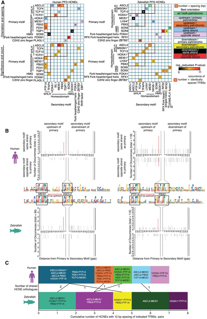
Consistent 12 bp spacing of homeodomain–bHLH TFBS pairs is significantly enriched for a small subset of orthologous HCNEs. (A) Matrix of TFBS motifs from Figure 5 and Supplemental Figure S19 showing consistent spacing in SpaMo analysis. Cells in the upper matrices are color-coded according to best orientiation, that is, the strand and position of the secondary motif relative to the primary motif for the most significant spacing. However, significant spacing may be present in other orientations also. Numbers in cells indicate the spacing of motifs in base pairs that is significantly enriched. Numbers in black indicate significant spacing in only one of human and zebrafish; gold numbers indicate motif pairs significant for HCNEs of both species. Cells in the lower matrices are color-coded based on significance of TFBS spacing. Numbers in cells indicate the number of occurrences of that spacing in significant orientations defined by SpaMo. Homeodomain–bHLH pairs are indicated in bold black boxes. (B) Distance and orientation plots of the two TFBS pairs identified as significant in both human and zebrafish HCNEs. Quadrants within individual plots are as labeled on the figure. Red lines within graphs indicate secondary TFBS locations contributing to significant spacing from the primary TFBS (depicted in the center), and gray bars indicate all other instances of the secondary TFBSs. (C) Bar graphs indicating cumulative numbers of HCNEs containing homeodomain–bHLH TFBS pairs exhibiting 12 bp spacing. The TFBSs pairs from A in which the TFBS pairs were identified are annotated on bar segments. The numbers of orthologous HCNEs shared between segments are indicated between the bars. The genomic locations of the six HCNEs indicated are shown in Supplemental Figure S22.