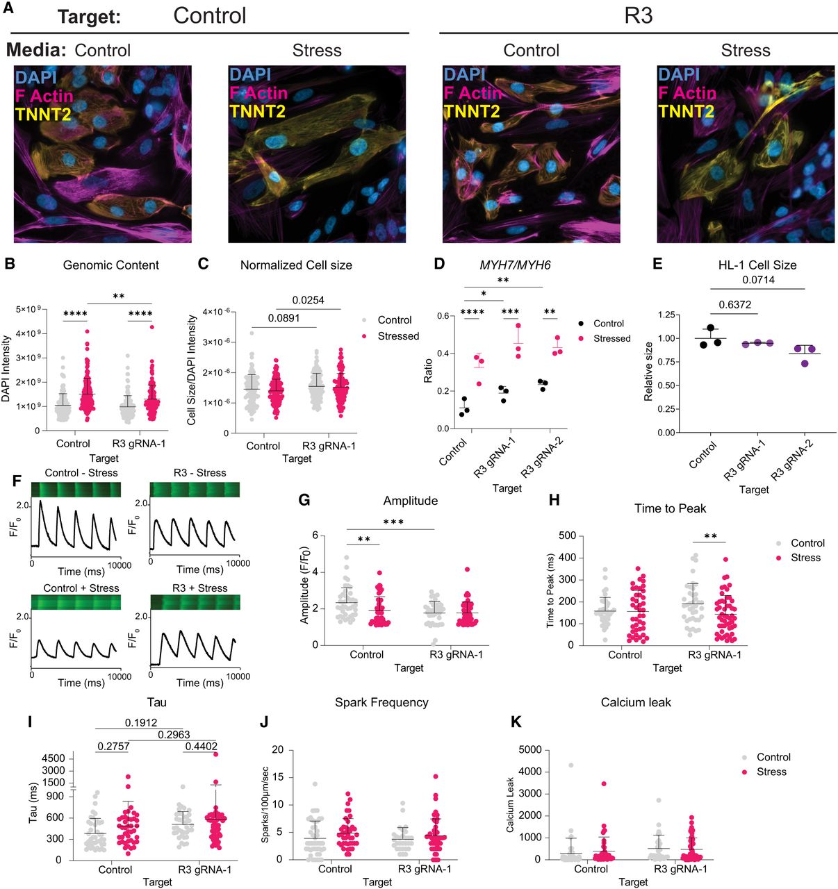
Activation of R3 by VP64dCas9VP64 (CRISPRa) alters cellular composition and function in control and stress states. (A) Representative images of iPSC-CMs across conditions. (B,C) Analysis of genomic content by DAPI intensity (B) and normalized cell size (C) of human iPSC-CMs represented in A 20 days following activation of each target ± ET-1 (1 µM) for the last 48 h of culture; n = 3 replicates. Statistics were calculated with a two-way ANOVA with Fisher's LSD. (D) The ratio of MYH7 to MYH6 abundance was measured using the data from Figure 3, F and G. Statistics were calculated on dCt values (normalized to TBP), and a two-way ANOVA with a Tukey's post hoc test was used to compare ratios. (E) Relative cell size measured 21 days following cCRE activation in HL-1 mouse atrial CMs (n = 3 replicates, mean ± SD). A one-way ANOVA test with a Dunnett's post hoc test was used to compare cell size. (F) Representative confocal line scan and Cal-520 traces in iPSC-CMs. (G–K) Quantitative analysis of cells represented in F (n = 6 replicates across two differentiations). Statistics were calculated with a two-way ANOVA with a Fisher's LSD. Calcium measurements are as follows: calcium amplitude (F/F0; G), time to peak (H), tau (I), spark frequency (J), and calcium leak (K). (*) Padj < 0.05, (**) Padj < 0.01, (***) Padj < 0.001, (****) Padj < 0.0001. See also Supplemental Figure S8.