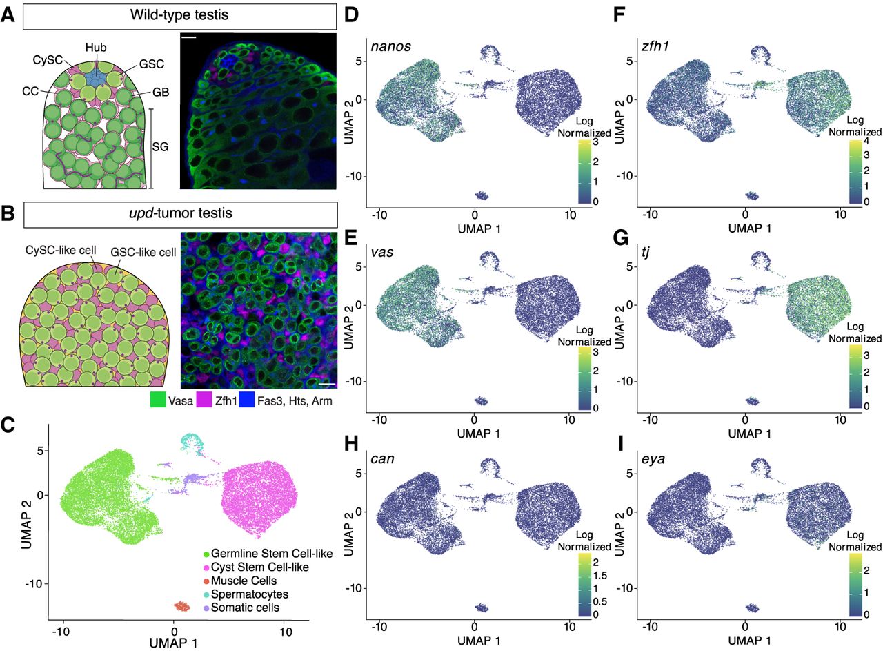
Single-cell RNA sequencing of upd tumor testes classifies two stem-cell populations. (A) Schematic of a wild-type testis alongside a single z-slice confocal image demonstrating the differentiation process of germline cells (green; Vasa-positive) and cyst cells (magenta; Zfh1-positive). At the apical tip, GSCs and CySCs surround the stem-cell niche, called the hub (Fas3; blue cell cluster). Individual spermatogonial (SG) cysts are identified by the spectrosome/fusome (Hts) and the extracellular adhesion protein, armadillo (Arm), both blue in the confocal image. Scale bar = 10 µm. (B) A schematic and single z-slice confocal image of the upd tumor. The tumor lacks a stem-cell niche and is composed of GSC-like (green; Vasa-positive) and CySC-like (magenta; Zfh1-positive) cells. In blue, is the cell adhesion protein armadillo (Arm) and the spectrosome (Hts), a spectrin-containing structure that appears as a dot in the cytoplasm of GSCs. The Vasa-positive, spectrosome-containing germline cells in the upd tumor display a morphology that is consistent with wild-type GSCs. Scale bar = 10 µm. (C) The upd tumor is composed of approximately five cell clusters, with the two largest groups representing GSC-like and CySC-like cells. (D–I) Uniform manifold approximation projection (UMAP) graphs show the cell- and stage-specific log-normalized expression of known marker transcripts that accurately define the GSC-like and CySC-like clusters.