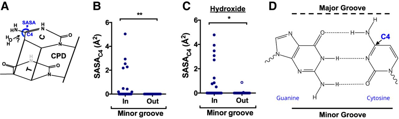
Molecular mechanism underlying elevated CPD deamination at minor-in rotational settings in nucleosomes. (A) Chemical structure of a thymine–cytosine CPD. Previous studies indicate that the rate-limiting step of cytosine deamination is hydrolytic attack by a water (H2O) molecule on the cytosine C4 position. The accessibility of these C4 position to hydrolytic attack can be quantified using the solvent accessible surface area (SASA). (B) Quantification of the SASAC4 for cytosine bases in the yeast nucleosome structure (1ID3) for bases at minor-in or minor-out rotational settings. (**) P < 0.01 based on a Mann–Whitney U test. (C) Same as panel B, except the SASAC4 is calculated for hydroxide (radius ∼ 1.1 Å) instead of water. (*) P < 0.05 based on a Mann–Whitney U test. (D) Chemical structure of the guanine–cytosine base pair, highlighting the fact that the cytosine C4 position is located near the major groove.