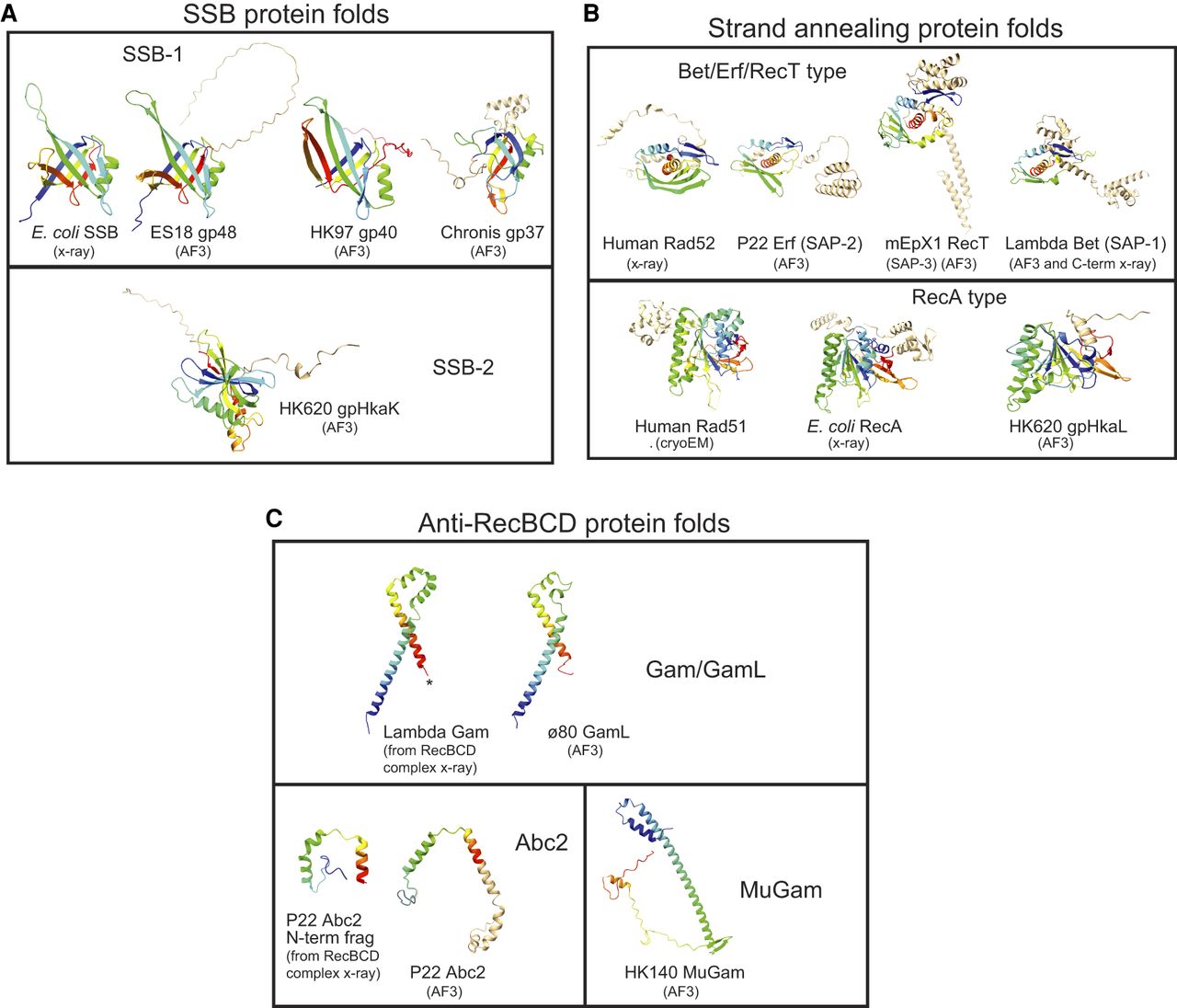
Polypeptide folds of Rec module proteins. Ribbon diagrams are shown for protein folds for single-strand binding proteins (SSBs; A), strand-annealing proteins (SAPs; B), and anti-RecBCD proteins (C). The diagrams were created with ChimeraX-1.5 (Pettersen et al. 2004) from proteins folded by AlphaFold 3 (AF3) (Abramson et al. 2024) unless otherwise indicated as determined by x-ray diffraction or cryo-electron microscopy. The core folds of proteins are shown in rainbow mode with blue at the N terminus and red at the C terminus. “Extra” polypeptide chain not in the common core fold is shown in tan. PDB accession numbers for experimentally determined structures shown are as follows: E. coli SSB, 1SRU and 4MZ9; λ Gam, 2UUZ and 5MBV; N-terminal fragment of P22 Abc2, 8B1T; C-terminal fragment of λ Beta, 7UJL and 6M9K; human Rad51, 5H1B; human Rad52, 8RIL; and E. coli RecA, 7JY9 and 2REB.