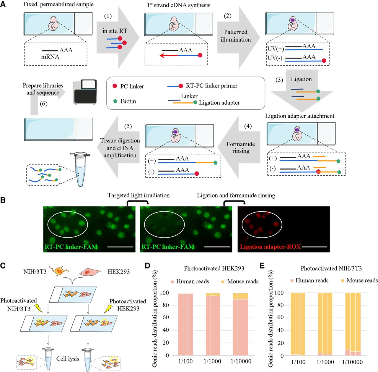
Overview of PCL-seq for ROI-specific expression analysis. (A) PCL-seq workflow: (1) Fixed and permeabilized tissue sections are subjected to in situ reverse transcription with RT primers carrying PC linkers, synthesizing the first strand of cDNA. (2) Patterned UV illumination of the ROI is performed using the Mosaic system, cleaving the PC linkers and exposing 5′ phosphate groups. (3) The cDNA is ligated to partially double-stranded adapters. The ligation adapter (top strand) is biotinylated at the 5′ end and consisted of three components: a high-throughput sequencing handle, a barcode, and a linker region complementary to the bottom strand. (4) High-concentration formamide washing removes unligated adapters, primarily from non-UV-illuminated regions. (5) After Proteinase K digestion to dissolve tissue, biotinylated cDNA is enriched using streptavidin-coated magnetic beads. A second reverse transcription step introduces the ISPCR sequence at the 3′ end, followed by cDNA amplification. (6) The amplified cDNA is fragmented using Tn5A transposase loaded with Illumina Nextera Read1 sequencing adapters, and sequencing libraries are prepared for downstream analysis. (B) The NIH/3T3 cell line is stained with RT-PC linker-FAM primers during the in situ RT reaction, followed by imaging after photocleavage, ligation, and formamide washing. The elliptical area indicates the illuminated region. Scale bar, 100 µm. (C) PCL-seq specificity is assessed by selectively photo-tagging HEK293 and NIH/3T3 cells in coculture. (D,E) Background signal levels from nonilluminated regions are evaluated in mixed human-mouse cell cultures (n = 3 replicates).