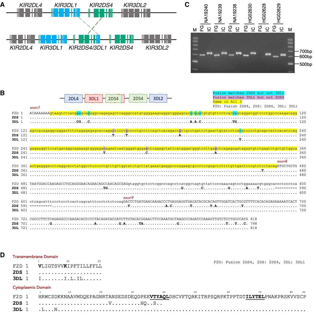
Identification of a novel fusion gene, KIR2DS4/3DL1. (A) A schematic diagram illustrating the formation of a novel KIR2DS4/3DL1 fusion gene composed of exons 1–7 from KIR2DS4 and exons 8–9 from KIR3DL1. (B) Sequence alignments comparing the intron 7 region of the fusion gene with corresponding regions in KIR2DS4 and KIR3DL1 for HG02630-M and NA19240-P. The highlighted yellow regions, signifying the sequences common to all three genes, are potential sites of the crossover event leading to the formation of the fusion gene. (C) An electrophoresis image displaying the results from the two trios of HG02630, NA19240, and their respective parents. Five out of the six samples contain the KIR2DS4/3DL1 fusion gene (indicated by FG), whereas all six samples carry the KIR3DL1 control gene as an internal control (indicated by IC). (D) The translated protein sequences of the KIR2DS4*00101, KIR3DL1*03501, and the KIR2DS4/3DL1 fusion genes were aligned to assess potential functional alterations in the fusion gene. Variations in the transmembrane domain are highlighted in bold. The transmembrane domain is encoded by the DNA sequence of exon 7, so the KIR2DS4/3DL1 fusion gene shares identical transmembrane domain residues with KIR2DS4*00101. The cytoplasmic domain, encoded by exons 7, 8, and 9, incorporates contributions from KIR3DL1*03501, including two inhibitory tyrosine-kinase motifs (ITIMs; I/VxYxxL/V), which are underscored and in bold in the alignment (Carr et al. 2005; Campbell and Purdy 2011; Oszmiana et al. 2016).