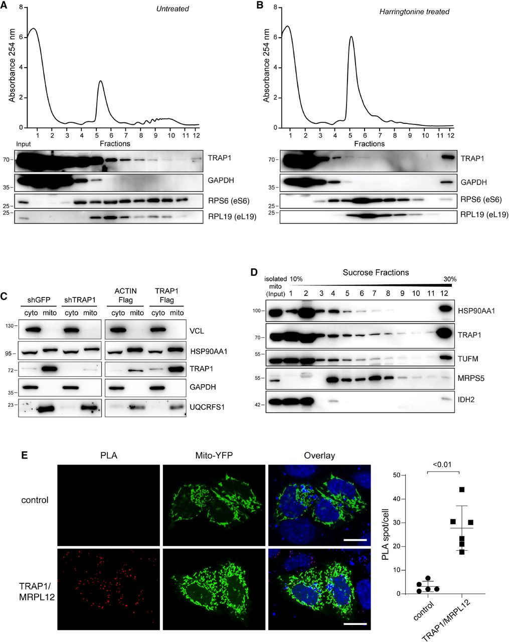
TRAP1 is associated with both cytosolic and mitochondrial ribosomes. (A,B) Polysome profiling absorbance, measured at 254 nm, of HeLa cell extracts, from untreated cells (A) or following a 5-min treatment with 2 µg/mL harringtonine (B). Fractions 1 and 2 are free cytosolic proteins or light complexes; fractions from 3 to 6, ribosomal subunits (60S, 40S) and monomer (80S); and fractions from 7 to 12, polysomes. Proteins from each fraction were analyzed by WB with the indicated antibodies. (C) Subcellular fractionation of HeLa cells showing the presence of indicated proteins into cytosolic (cyto) and mitochondrial (mito) fractions. VCL and GAPDH have been used as markers of cytosol and UQCRFS1 as a marker of mitochondria. (D) HeLa cell mitochondria were isolated, lysed, and loaded onto a 10%–30% linear sucrose gradient, followed by fractionation. Proteins were precipitated from the resulting fractions and subjected to western blot with indicated antibodies. (E) Representative image of PLA showing the interaction between TRAP1 and MRPL12 in HeLa mitochondria. Positive signals of interaction are shown as red dots; nuclei are stained with DAPI (blue); and mitochondria are marked by the mitochondria-directed YFP (green). The control PLA has been obtained by hybridizing with anti-TRAP1 only as primary antibody. Scale bar, 10 µm. The graph shows the average number of PLA spot/cell, with a P-value representing the statistical significance based on the Student's t-test (n = 6).