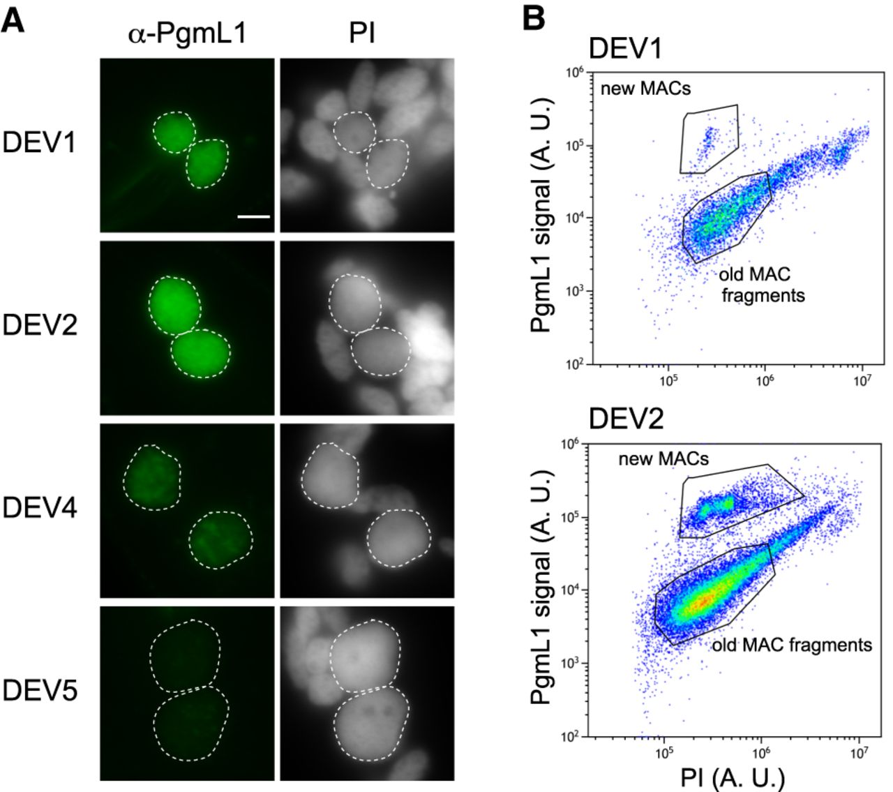
Figure 1.
PgmL1 immunostaining during autogamy. (A) Whole-cell immunostaining at different stages of autogamy time course 1 (tc1). New MACs and fragments are counterstained with propidium iodide (PI). Developing MACs are surrounded by a white dotted line. Scale bar is 5 µm. Developmental stages (DEV1 to DEV5) are defined in the Methods. (B) Flow cytometry analysis of immunostained nuclei at the DEV1 and DEV2 stages of autogamy time course 2 (tc2). Following gating of total nuclei (see Supplemental Fig. S1D), the population of new MACs was separated based on their PgmL1 signal. The PI axis is indicative of DNA content. (A. U.) Arbitrary units in log scale.