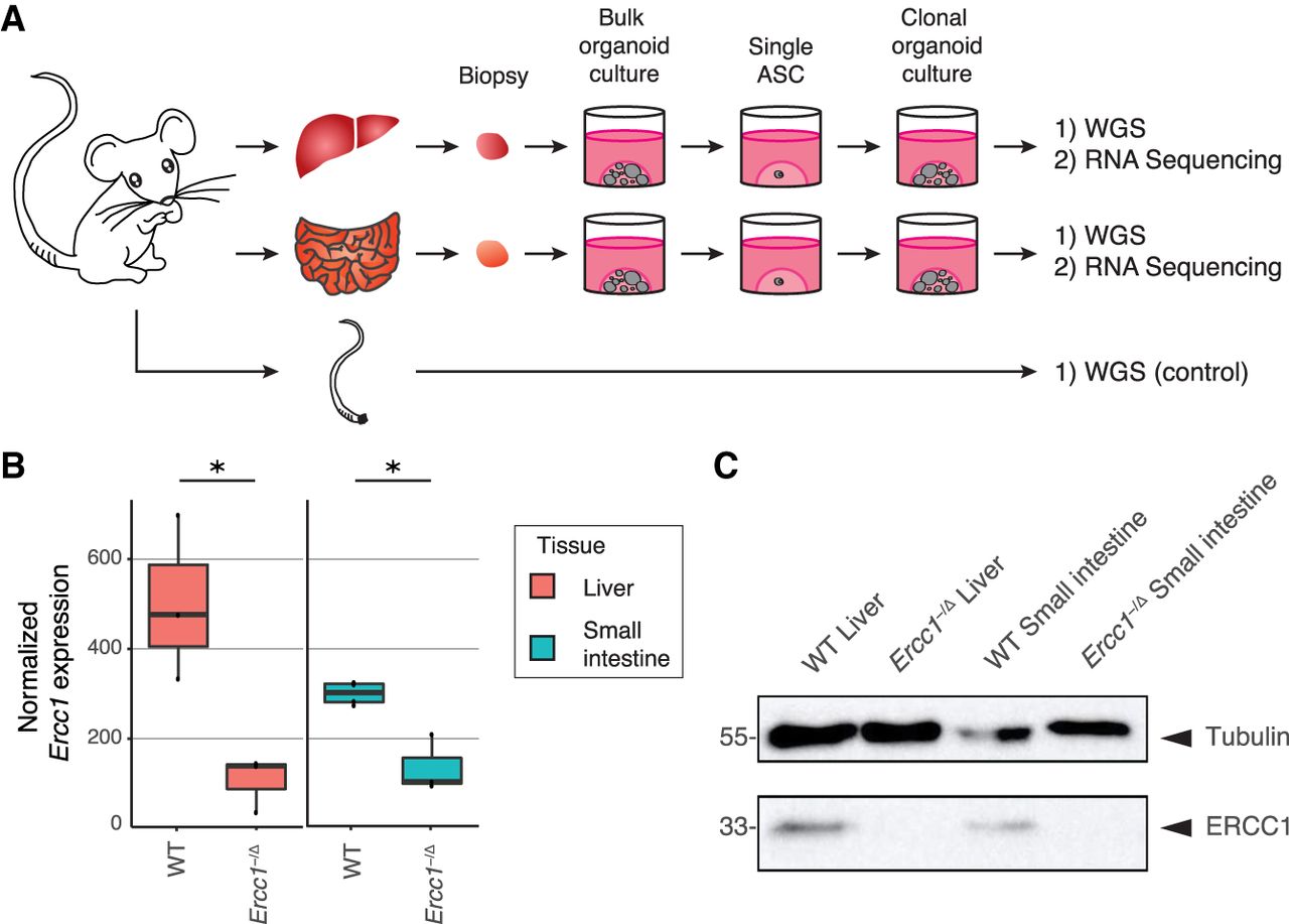
Experimental setup and tissue-specific expression of Ercc1 in mouse ASCs. (A) Schematic overview of the experimental setup used to determine the mutational patterns in single ASCs from the liver and small intestine of mice. Biopsies from the liver and small intestine of six 15-wk-old female mice (three Ercc1−/Δ mice and three WT littermates) were cultured in bulk for ∼1.5 wk to enrich for ASCs. Subsequently, clonal organoids were derived from these bulk organoid cultures and expanded for ∼1 mo, until there were enough cells to perform both WGS and RNA sequencing. As a control sample a biopsy of the tail of each mouse was also subjected to WGS. (B) Box plots depicting normalized Ercc1 expression in ASC organoid cultures derived from liver and small intestine of Ercc1−/Δ mice (n = 3 and n = 3, respectively) and WT littermates (n = 3 and n = 4, respectively). Asterisks represent significant differences (P < 0.05, negative binomial test). (C) Western blot analysis of ERCC1 in Ercc1−/Δ and WT small intestinal and liver mouse organoids.