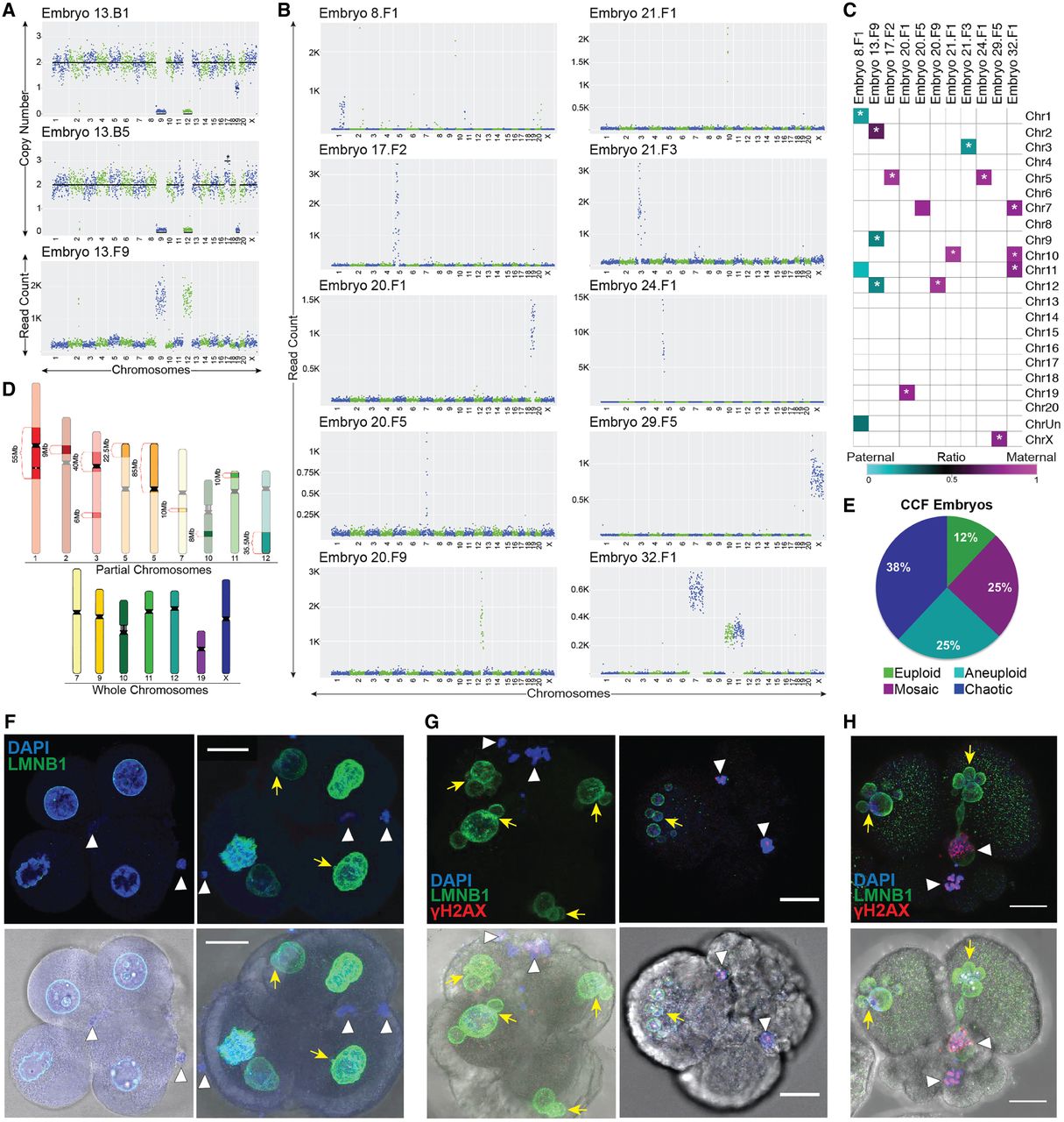
Chromosomes are eliminated via cellular fragmentation and are susceptible to DNA damage. (A) CNV or read count plots demonstrating that Chr 9 and Chr 12 lost from two blastomeres (top and middle; also missing one to two copies of Chr 19) were detected in a cellular fragment (bottom) from the same embryo. (B) Additional examples of individual, multiple, and/or partial chromosomes in fragments of rhesus embryos. (C) Heat map of maternal versus paternal SNP genotyping ratios showing that CCFs can originate from the mother or father. White asterisk demarcates significant P-values (P < 9.1 × 10⁻⁶) for cumulative binomial test with Bonferroni correction. (D) Rhesus ideograms representing whole (bottom) and partial (top) chromosomes with approximate sizes highlighted that were detected in fragments. (E) Percentage of embryos with CCFs (N = 8 embryos) that were chaotic (blue), aneuploid (turquoise), mosaic (magenta), and euploid (green). (F) Five-cell embryos with normal-appearing blastomeres containing micronuclei (yellow arrows) and CCFs (white arrowheads) identified by DAPI (blue) and LMNB1 (green). Brightfield images (bottom) provided for reference. (G) Other cleavage-stage embryos with multiple micronuclei and CCFs also immunostained for the double-stranded DNA break marker, γH2A.X (red), showed that the chromosomes within fragments are unstable and damaged. (H) One embryo also contained a micronucleus that appeared to be in the process of CCF sequestration. Scale bar, 25 µm.