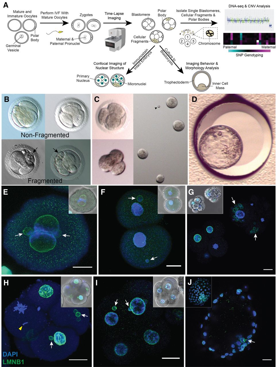
Approach for assessing micronuclei, fragmentation, and CNV in rhesus embryos. (A) Mature and immature oocytes were obtained from female rhesus macaques undergoing controlled ovarian stimulations. MII oocytes displaying one polar body were fertilized by conventional IVF with sperm from rhesus males. (B) Early mitotic divisions and the incidence of cellular fragmentation in presumptive zygotes (identified by two pronuclei and/or polar bodies) were analyzed by time-lapse imaging. (C) Cleavage-stage embryos were disassembled into individual blastomeres, cellular fragments, and polar bodies for CNV and SNP analysis by scDNA-seq (N = 50). A subset of intact cleavage-stage embryos was fixed and immunostained for confocal imaging (N = 25). (D) Another group of embryos were cultured up to the blastocyst stage (N = 92). (E) Immunostaining of a zygote undergoing syngamy with two micronuclei (white arrows) shown by the nuclear envelope marker, LMNB1 (green); DAPI (blue) = DNA. (F) Two-cell embryo with one micronucleus in each blastomere. (G) Comparison of a fragmented (white arrowheads) cleavage-stage embryo with multiple micronuclei (right) and a nonfragmented seven-cell embryo (left). (H) Single imaging plane of a Z-stacked five-cell embryo with a missegregated chromosome (yellow arrowhead) and micronuclei, as well as a (I) nine-cell embryo showing micronuclei in two blastomeres, but no visible cellular fragmentation. Insets show a brightfield image for reference. Scale bars, 25 µm. (J) Blastocyst with two micronuclei in the ICM; the inset shows the maximum intensity projection of the embryo.