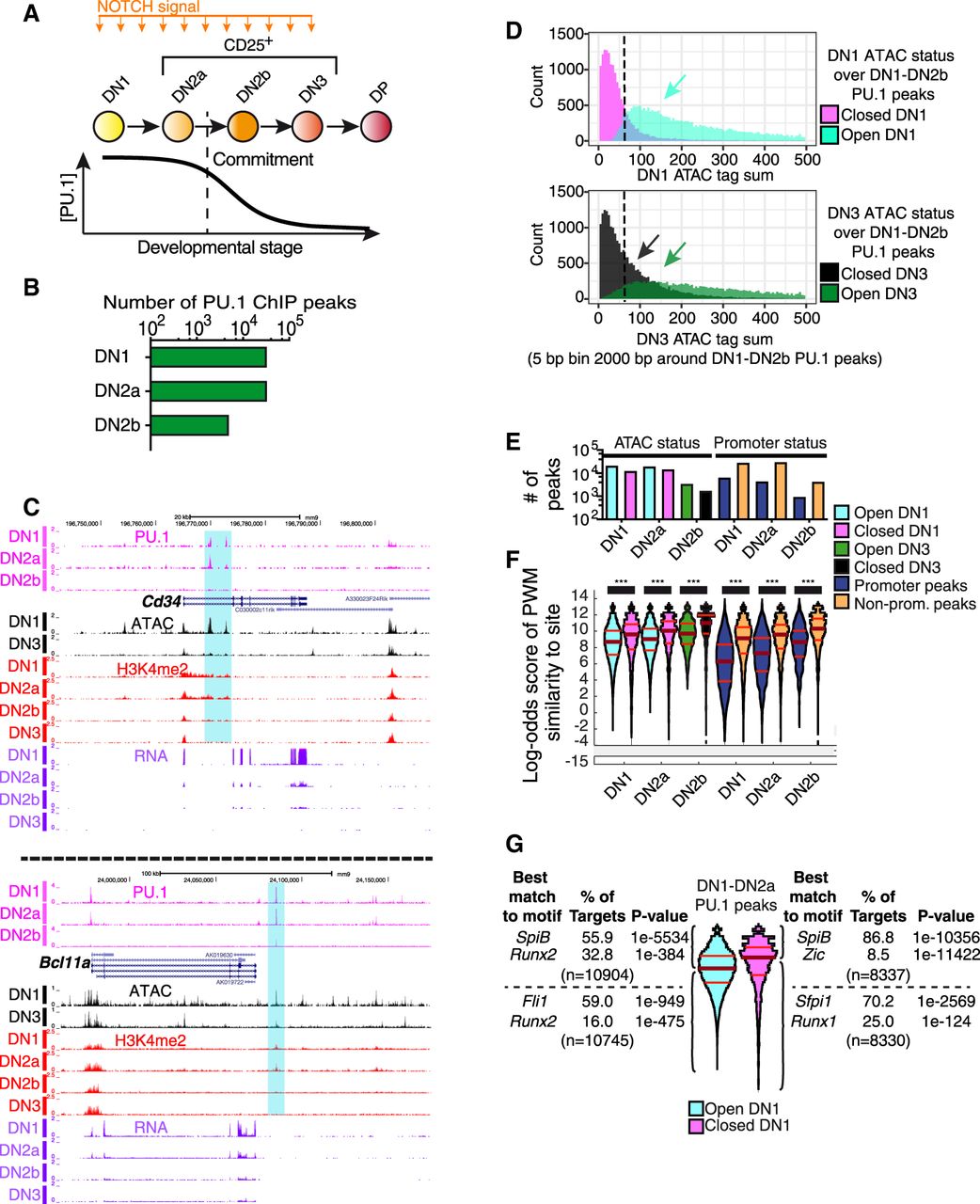
Endogenous PU.1 binding in pro-T cells: distinct affinity thresholds for binding at open and closed sites and promoter and nonpromoter sites. (A) Outline of early T-cell development with schematic depicting PU.1 expression levels. (DP) A later stage. (B) Number of endogenous PU.1-occupancy peaks detected in DN1, DN2a, and DN2b pro-T cells. (C) Cd34 and Bcl11a UCSC Genome Browser tracks (http://genome.ucsc.edu) showing endogenous PU.1 ChIP, ATAC-seq, H3K4me2 ChIP, and RNA-seq in the DN1, DN2a, DN2b, and/or DN3 stages. Samples from in vitro differentiation from fetal liver precursors or from thymus (ATAC, DN1, DN3). Data from GSE31235; GSE93755. (D) Histogram of ATAC tag counts at DN1 (top) or DN3 (bottom) stages across all pro-T-cell PU.1 binding sites. Sites in regions defined as ATAC “open” or “closed” (Methods) are plotted separately to aid visualization. (Y-axes) Number of sites at indicated ATAC signal level. (E) Number of PU.1 peaks in indicated stages in ATAC-open and ATAC-closed as well as promoter and nonpromoter regions. The same color key is used in E and F. (F) Distribution of PU.1 motif log-odds scores at binding sites in open (cyan: open in DN1; green: open in DN3) and closed regions (magenta: closed in DN1; black: closed in DN3), and in promoters (dark blue) and nonpromoter (orange) elements. Scores from a DN1-DN2b-derived PU.1 PWM-matrix (Supplemental Table S1). Kruskal–Wallis statistical test: (***) P ≤ 0.0001. (G) Motif analyses of PU.1 sites in DN1-DN2a cells, classified by PWM scoring and ATAC accessibility in DN1.