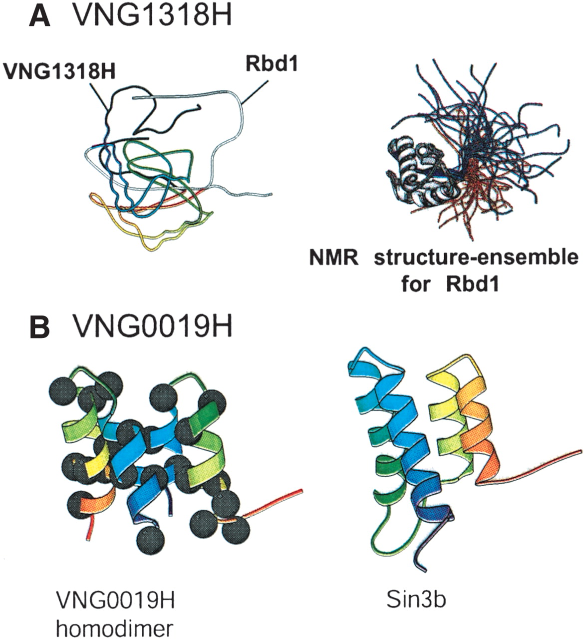
Figure 5
Functional annotation of proteins through ab inito structure prediction by Rosetta. (A) Alignment of the Rosetta-predicted structure for VNG1318H and Rbd1 (left). The NMR structure for Rbd1 is shown on the right. (B) Rosetta-predicted structure for the VNG0019H homodimer (left) and its closest match, the NMR structure for Sin3b. The black dots indicate hydrophobic residues located primarily at the predicted protein–protein interface.