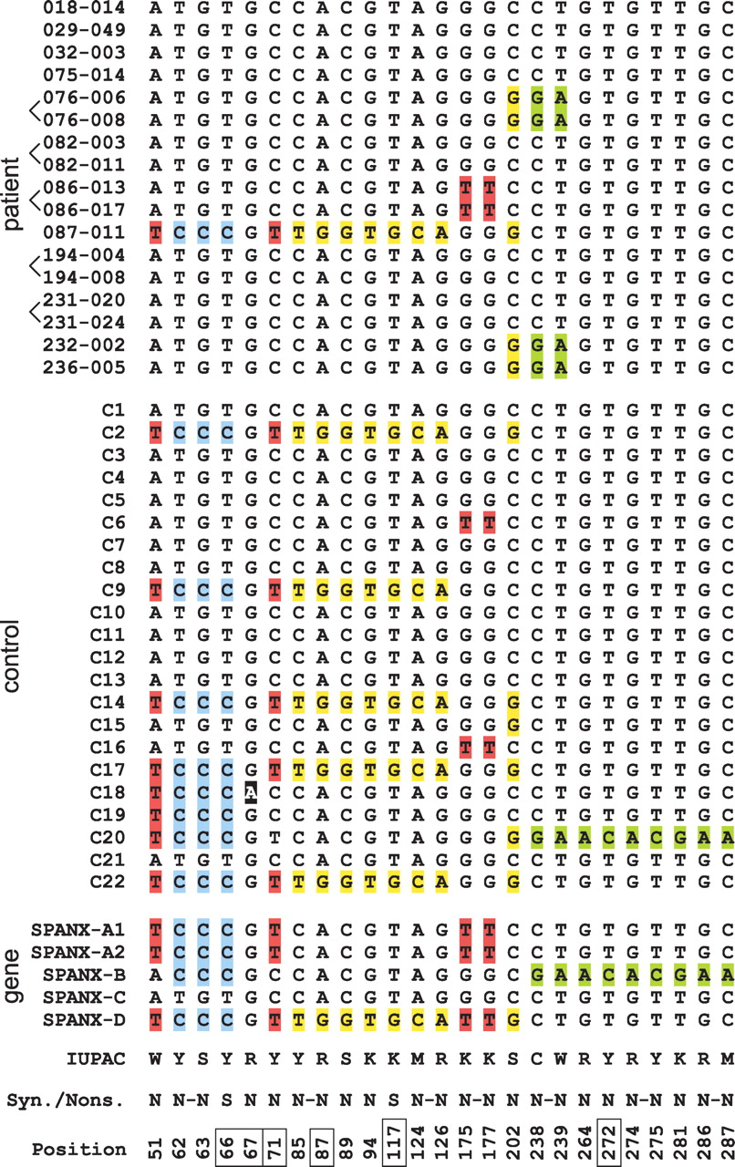
Analysis of SPANX-C coding sequences in X-linked families. The alignment of the SPANX-C coding sequences obtained from prostate cancer patients (top), healthy controls (middle), and genomic SPANX-A/D genes (bottom). The figure shows the results of analysis of 12 families, some of them are represented by two brothers (brackets show brothers). Note that position 67 (black shading) is a single point mutation without gene conversion; all other mutation positions are recombinations/gene conversions. IUPAC codes of the variants, synonymous (S) and nonsynonymous (N) characters of changes, and positions in the CDS alignment are shown at the bottom; positions highlighted by boxes correspond to CpG sites and their TpG/CpA variants. If several mutations were in the same codon, they were marked by a dash. In all such cases, the resulting effect was an amino acid replacement, and therefore, all such changes were marked as nonsynonymous.