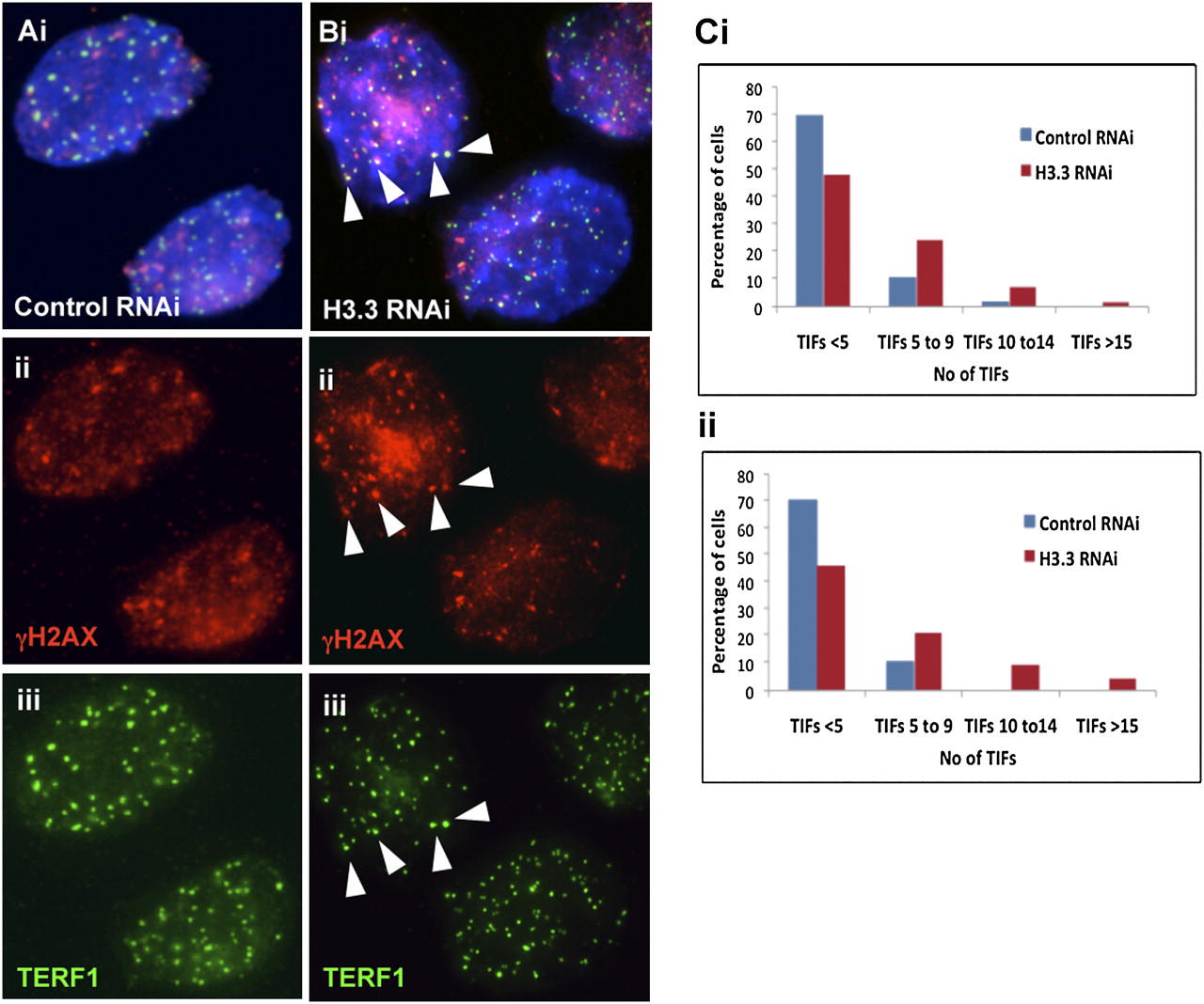
Telomere damage response upon H3.3-depletion. (A,B) Immunofluorescence analysis of ES129.1 cells subjected to 48-h knockdown with either control or H3.3-specific RNAi-duplex oligonuclotides using anti-gamma-H2AX (A,B, ii, red) antiserum. Increased number of TIFs was detected at the telomeres (indicated by colocalization with TERF1; A,B, iii, green) in cells depleted of endogenous H3.3 (B) (arrowheads show some examples of TIFs). (C) Induction of TIFs by H3.3 inhibition for 48 h. Data are presented in histograms by subgrouping the cells according to the number of TIFs per cell (<5 TIFs, 5–9 TIFs, 10–14 TIFs, and ≥15 TIFS). A normal cell can contain 1–2 TIFs on average, thus, a threshold of 5 TIFs was used, as described in other studies (Hockemeyer et al. 2005). When transfected with H3.3-RNAi oligonuclotides, the number of cells with ≥5 TIFs increased from 12.94% to 37.65% (n = 85; 24.71% increase; P = 3.80 × 10⁻⁵) and 11.76% to 40.0% (n = 85; 28.24% increase; P = 6.26 × 10⁻⁵) for the two experiments (i and ii), respectively, with an average increase of 26.47%, compared with the population transfected with control scrambled RNAi-oligonucleotide. Data for 24-h H3.3 depletion in ES129.1 and NIH3T3 cells are shown in Supplemental Figure 13.