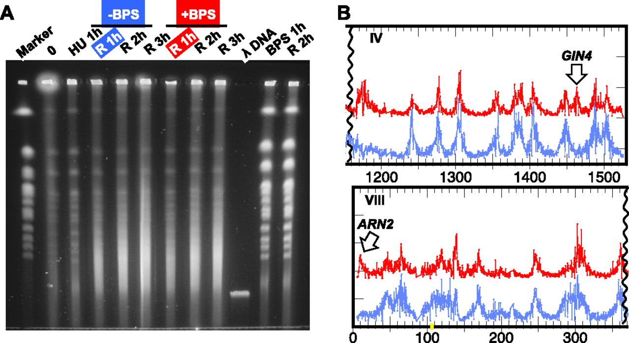
Figure 5.
Treatment with BPS induces additional breakage at discrete loci. (A) Chromosome breakage is readily observed in mec1 cells recovering from HU in the absence (blue, −BPS) and presence of 0.8 μM BPS (red, +BPS) by PFGE. BPS addition alone does not cause chromosome breakage: “BPS 1h” and “R 2h” represent cells that have been treated with BPS for 1 h and those that have recovered in fresh medium for 1 h after treatment with BPS, respectively. (Marker) Yeast chromosome PFG marker; (λ DNA) phage lambda DNA, ∼50 kb. (B) Break-chip demonstrates examples of genomic regions showing enhanced breakage in the +BPS sample (red) compared to the –BPS sample (blue), as indicated by arrows.