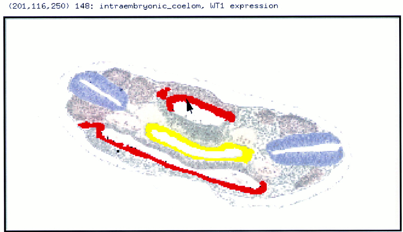
Figure 3.
Gene expression data. A coronal section through the body region of an E14 embryo showing WT1 expression (red, data from Armstrong et al. 1992) in the lateral wall of the coelomic cavity (the presumptive mesothelium) and in the intermediate mesoderm (which has yet to segregate into nephric duct and cord—cf. with Fig. 4). Other tissues highlighted are the gut (yellow), the neural tube (blue), the somites (light brown), and the dorral aortae (very pale pink).