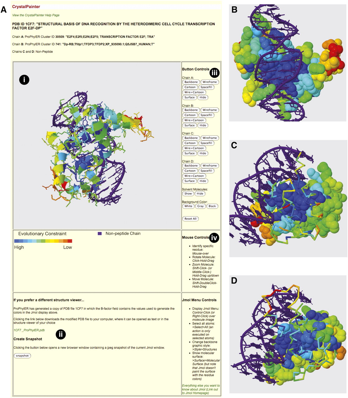
CrystalPainter. (A) A screenshot of the CrystalPainter page for PDB ID 1CF7. Major features include: (i) the Jmol window, where the molecular image is manipulated; (ii) links to download a coded structure file or an image of the Jmol window; (iii) button controls to modify the display settings; and (iv) instructions for mouse controls and navigating the Jmol window. (B) Image capture from CrystalPainter. The peptide chains have been displayed as “Spacefill” to highlight the conserved residues on the surface (shades of blue) and the structure reoriented to view the DNA-binding surface. (C) Chain A has been displayed as “Backbone” and Chain B as “Spacefill,” and the structure reoriented to better view the dimerization surface of Chain B. (D) Chain B has been displayed as “Backbone” and Chain A as “Spacefill,” and the structure reoriented to better view the dimerization surface of Chain A.