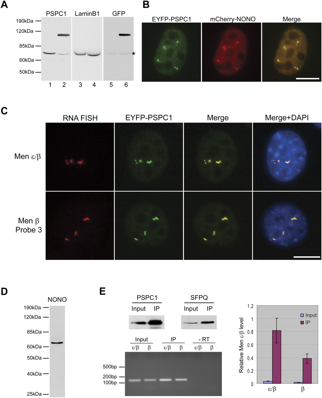
Men ε/β transcripts are localized to nuclear paraspeckles. (A) A C2C12 cell line stably expressing EYFP fused to PSPC1 (also known as PSP1α) was established. Immunoblotting showed that the endogenous PSPC1 level was reduced in (lane 2, lower band) C2C12 EYFP-PSPC1 stable cells compared to in (lane 1) wt C2C12 cells. (Lanes 3,4) Lamin B1 served as a loading control in a duplicate blot. (Lanes 5,6) After stripping the anti-Lamin B1 antibody, immunoblotting using an anti-GFP antibody confirmed that the band at ∼100 kDa corresponds to EYFP-PSPC1. (*) Residual Lamin B1 signal. (B) mCherry fused to NONO (also known as p54/nrb) was transiently expressed in C2C12 EYFP-PSPC1 stable cells. The foci of mCherry-NONO are colocalized with EYFP-PSPC1. Scale bar, 10 μm. (C) RNA FISH analysis showed that the Men ε/β transcripts are localized to paraspeckles. A probe that detected both the Men ε and Men β transcripts, as well as a probe that only detects Men β, exhibit the same localization patterns. Scale bar, 10 μm. (D) A mouse monoclonal antibody to NONO, designated 9-99, was generated. Immunoblotting analysis using C2C12 whole-cell lysate detected a single band confirming the specificity of the 9-99 monoclonal antibody. (E) A coimmunoprecipitation assay revealed that the Men ε/β transcripts directly interact with NONO. Immunoblotting using anti-PSPC1 or anti-SFPQ antibodies after co-IP showed that PSPC1 and SFPQ (also known as Psf) directly interact with NONO. Ten percent input was used for immunoblotting. cDNA was generated using random hexamers from the IP fraction. RT-PCR revealed the existence of the Men ε/β transcripts in the NONO protein complex. Both Men ε/β transcripts are ∼20-fold enriched in the same IP fraction, assessed by Q-PCR. Gapdh was used as a normalization control in Q-PCR. The data in the histogram are shown as mean and standard deviation values of three technical replicates.