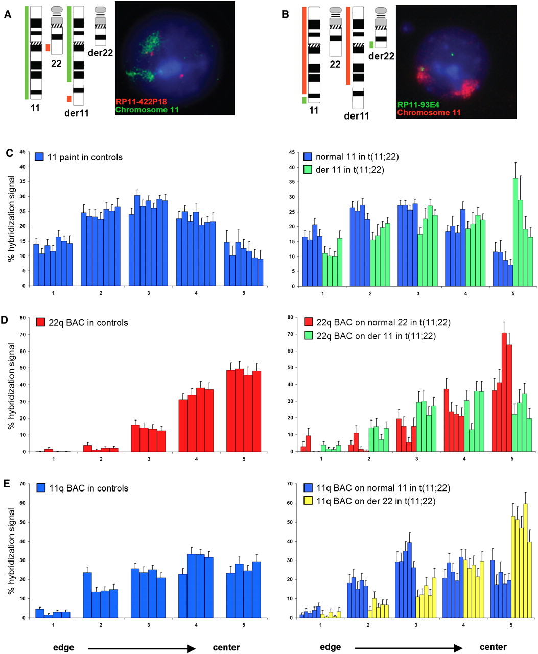
Nuclear position of normal and derivative chromosome territories in t(11;22) cells and control cell lines. BAC coordinates are shown in Table 1. (A) Chromosome ideograms and schematic representation of the regions recognized by FISH probes used to distinguish the normal and derivative chromosome 11s, and an example of FISH on lymphoblastoid cells from a t(11;22) carrier. (Green) Chromosome 11 paint; (red) BAC RP11-422P18. DNA is counterstained with DAPI (blue). In nuclei of cells carrying the balanced t(11;22), the normal HSA11 is marked solely by the green chromosome paint signal, whereas the green painted territory for the derivative chromosome 11 is also associated with a red BAC hybridization signal. The normal HSA22 is indicated by a lone red BAC signal. (B) As in A, but chromosome 11 paint is in red and BAC RP11-93E4 is in green. In nuclei of cells carrying the balanced t(11;22), the normal HSA11 is marked by the red chromosome paint signal associated with a green BAC hybridization signal, whereas the derivative chromosome 11 is marked solely by the red painted territory. The derivative chromosome 22 is indicated by a lone green BAC signal. (C) Mean (+ SEM) percent of chromosome 11 paint hybridization signal present in five shells of equal area eroded from the edge (shell 1) to the center (shell 5) of the nucleus in seven control cell lines of normal karyotype (left) and four t(11;22) cell lines (right), showing the relative positions of the normal (blue) and derivative chromosome 11 (green). n = 40–50 nuclei each for each cell lines. The derivative 11 territory is shifted to a more central position in the nucleus compared with the normal 11. (D) As in C, but using BAC RP11-422P18 on four normal cell lines (left) and four t(11;22) cell lines (right) to compare positioning of the normal chromosome 22 (red) from that of the derivative 11 (green). The BAC signal on the derivative 11 is situated in a more peripheral position than that on the normal chromosome 22. (E) As in C, but using BAC RP11-93E4 on four normal cell lines (left) and five t(11;22) cell lines (right) to compare positioning of the normal chromosome 11 (blue) from that of the derivative 22 (yellow). The BAC signal on the derivative 22 is situated in a more central position than that on the normal chromosome 11.