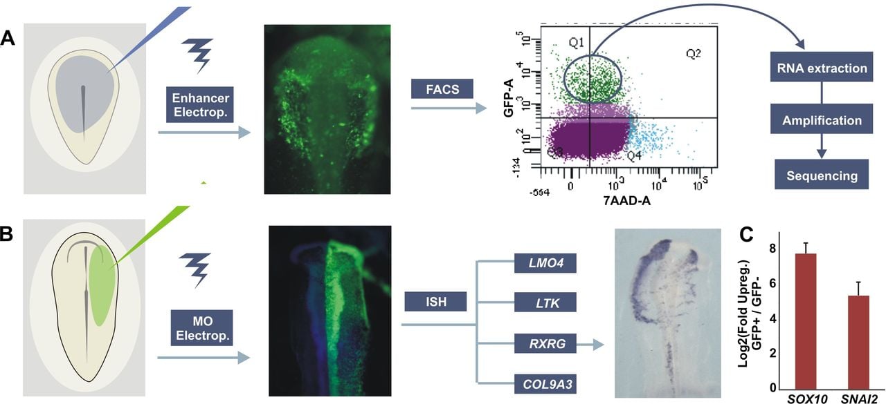
Experimental design for transcriptome analysis of cranial neural crest cells and loss-of-function studies. (A) Gastrula stage embryos were electroporated with Sox10e2:eGFP construct and incubated until HH10. In preparation for FACS, embryos were dissected and enzymatically dissociated. GFP+ cells were sorted with 7-AAD exclusion to eliminate damaged or dead cells. RNA from GFP+ and GFP− cells was extracted, reverse transcribed, and linearly amplified for high-throughput sequencing. (B) Loss-of-function studies in chicken embryos. Translation-blocking morpholinos (MO) targeted to known neural crest regulators were injected into one side of HH6 embryos, with the uninjected side used as an internal control. Electroporated embryos were allowed to develop until HH10 and expression of novel neural crest specific genes were analyzed by in situ hybridization (ISH). (C) qPCR confirm up-regulation of neural crest markers in sorted Sox10E2:eGFP+ cells in relation to GFP− cells.