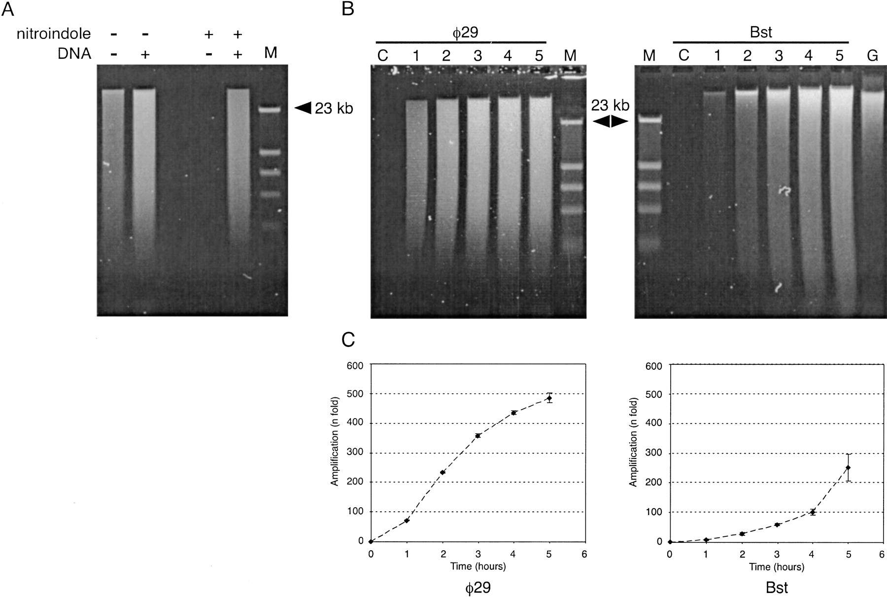
Gel electrophoresis analysis of amplified DNA. (A) Control reactions were incubated for 5 h in a 30-μL volume containing no DNA, or 7.5 ng of human DNA. Samples representing 5% of the reaction were denatured in alkaline buffer and analyzed on a 0.5% alkaline agarose gel, and stained with SYBR-green II (Molecular Probes). Lanes labeled M contained phage λ DNA digested with restriction endonucleaseHindIII. Reactions catalyzed by φ29 polymerase were incubated with (+) or without (−) input of denatured human DNA. Random heptamers contained standard (−) DNA, or were modified by the addition of two nitroindole groups (+) at the 5′ end. (B) Time-course reactions for φ29 and Bst DNA polymerases. Reactions were performed using nitroindole-modified primers. Every hour (from 1–5 h), 1.5 μL was removed, denatured in alkaline buffer, and analyzed in 0.5% alkaline agarose gel. Lanes labeled C correspond to control samples incubated for 5 h without input DNA. The lane labeled G corresponds to a gel load of genomic DNA equivalent to 100× the original DNA input of the amplification reactions. (C) Plots under gel images display the time course (fold amplification vs. time) of both polymerase reactions, generated by quantification of DNA yield with the PicoGreen Quantitation Kit. Background fluorescence at time 0 was subtracted for all time points. Each point represents the mean (±1 SD) of four independent analyses.