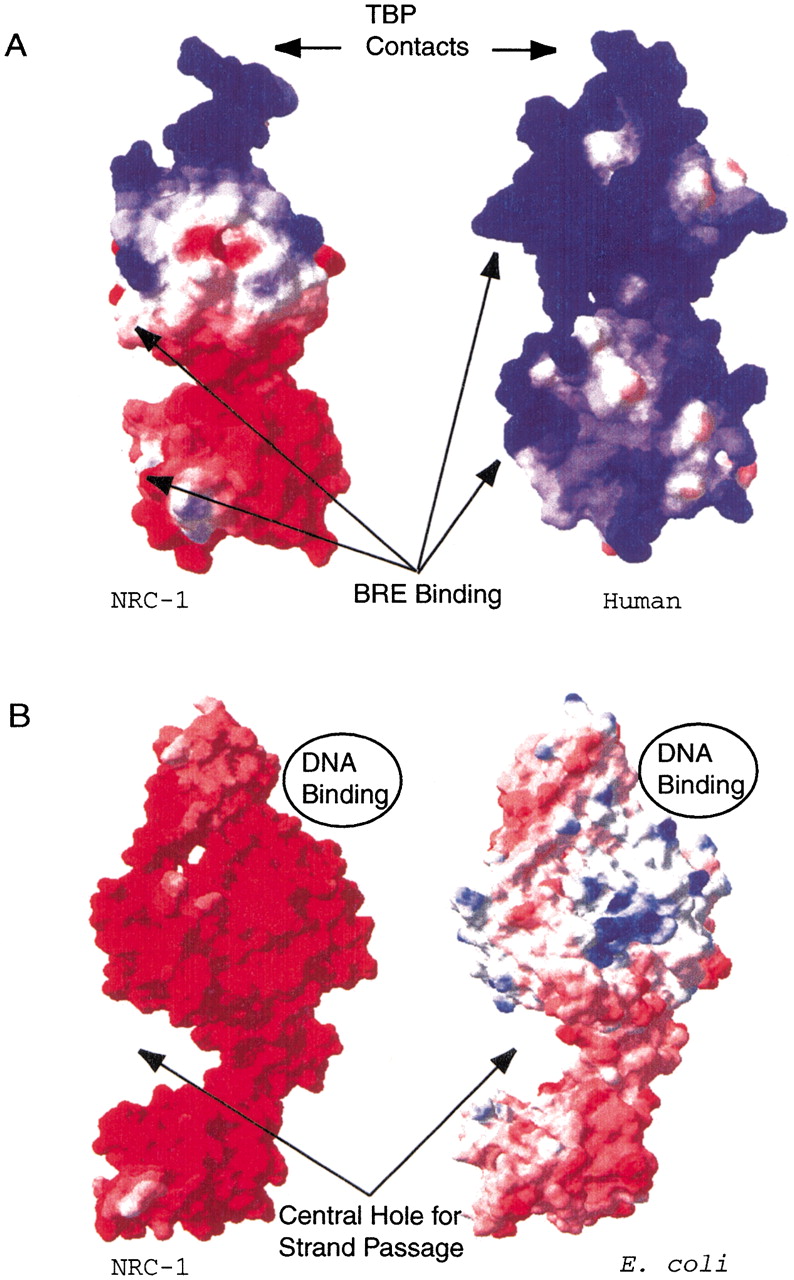
Figure 2.
Surface charge comparisons for halophilic and nonhalophilic proteins. Acidic character is indicated by red, basic character is indicated by blue, and neutral areas are indicated by white. (A) NRC-1 TFBe (left) and Human TFIIB (right) are shown with sites for BRE and TBP contacts indicated. (B) NRC-1 (left) and Escherichia coli GyrA (right) are shown with the binding site for the helix that is cleaved as well as the site for strand passage indicated for both molecules.