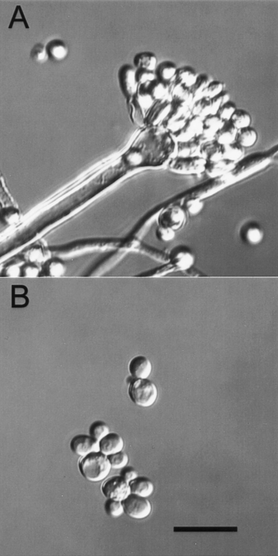
Figure 1.
Differential contrast micrographs of A. nidulans showing a condiophore, hyphae, and conidia (A) and S. cerevisiae cells (B). These micrographs were taken at the same magnification and illustrate how filamentous fungi produce a variety of complex cell types. Bar, (bottom panel), 10 μm.