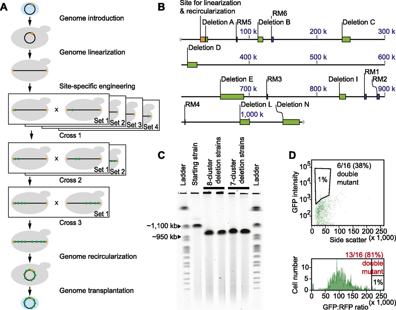
Progressive clustering of deleted genomic segments. (A) Scheme of the method. Light blue oval represents a bacterial cell. Black ring or horizontal line denotes a bacterial genome, with the orange box indicating the yeast vector used as a site for linearization and recircularization. Gray shape denotes a yeast cell. Green dot in the genome indicates a deletion replaced with a GFP marker. (B) Map of deleted regions. Orange box indicates the yeast vector sequence used for genome linearization and recircularization. Green boxes indicate regions deleted in multimutant mycoplasma strains. Blue boxes denote restriction modification (RM) systems that are also deleted in the strains. (C) Pulsed-gel electrophoresis result for deleted genomes. The starting strain was the JCVI-syn1.0 ∆1–6 strain (1062 kb). Two strains were analyzed for each design of simultaneous deletion (962 kb for eight-deletion or 974 kb for seven-deletion genome). Ladder is a set of yeast chromosomes (New England BioLabs). (D) GFP-RFP ratio sorting result. Standard sorting was compared with sorting based on a GFP-RFP ratio (Methods).