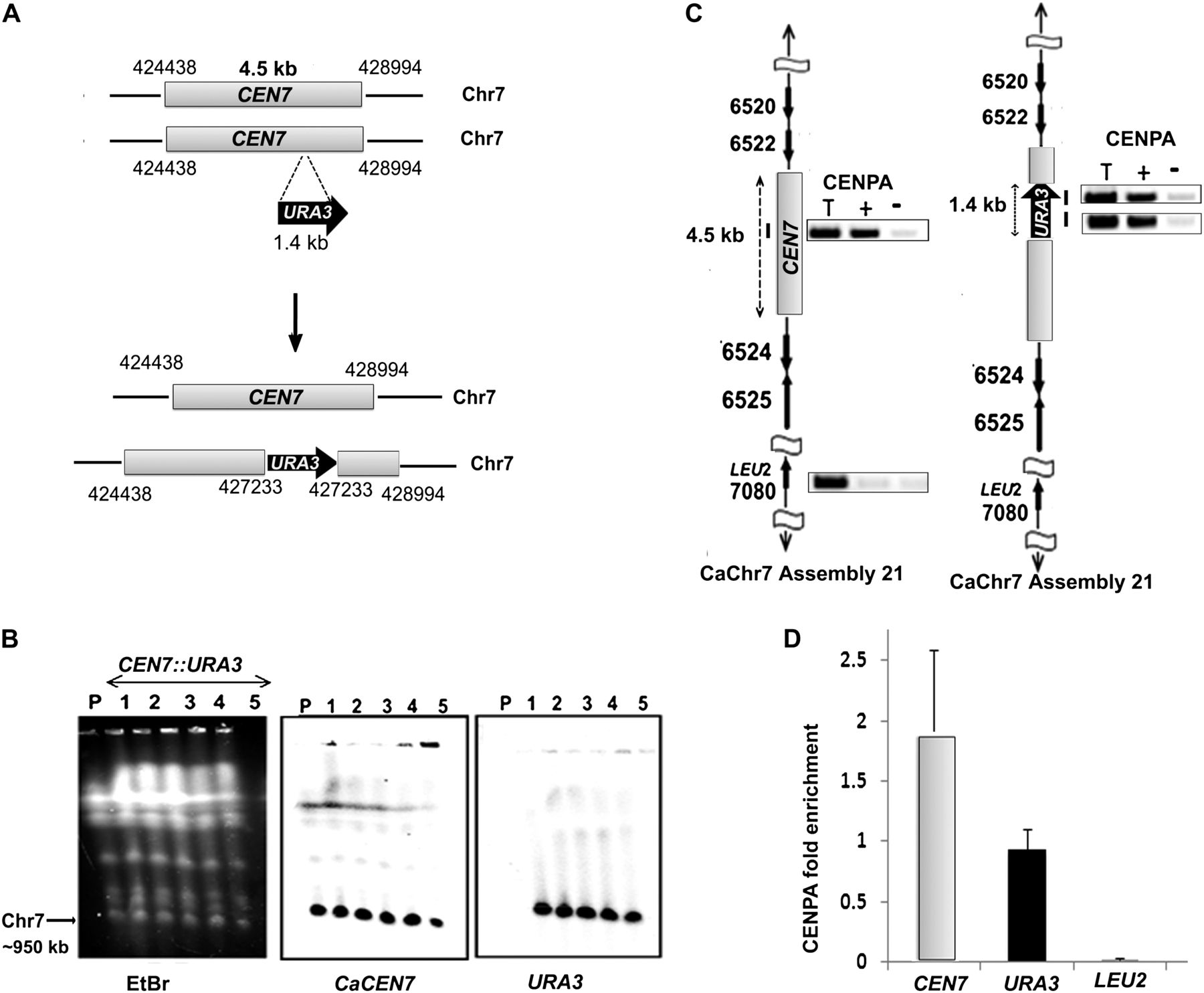
CENPA chromatin assembles at a specific chromosomal location. (A) Schematic showing the location of URA3 insertion at native CEN7 in one homolog of Chr7. Arrows indicate the gene location and the direction of transcription while the numbers represent coordinates on a specific chromosome based on Assembly 21 of C. albicans SC5314 genome. (B) An ethidium bromide (EtBr) stained CHEF gel image (left) showing no apparent alteration in the karyotype of RM1000AH-CEN7∷URA3 integrants (lanes 1–5) as compared with the parent RM1000AH (lane P). DNA was transferred on a membrane and Southern analysis was performed by probing the blot with either CaCEN7 (middle) or URA3 (right). (C) ChIP analysis with anti-Cse4 (CENPA) antibodies followed by PCR revealed recruitment of CENPA on URA3 of the altered homolog (left) as well as on native CEN7 of the unaltered homolog (right) of Chr7. Primer locations are marked with bars. LEU2 present on Chr7 but unlinked to CEN7 is used as a negative control for CENPA binding. (T) Total DNA; (+) with Ab; (−) without Ab (beads only). (D) Real-time qPCR analysis on CENPA ChIP DNA from RM1000AH-CEN7∷URA3/CEN7 strain J154 using primers from CEN7 and URA3 regions. Fold enrichment of CENPA binding was calculated as described in Methods.