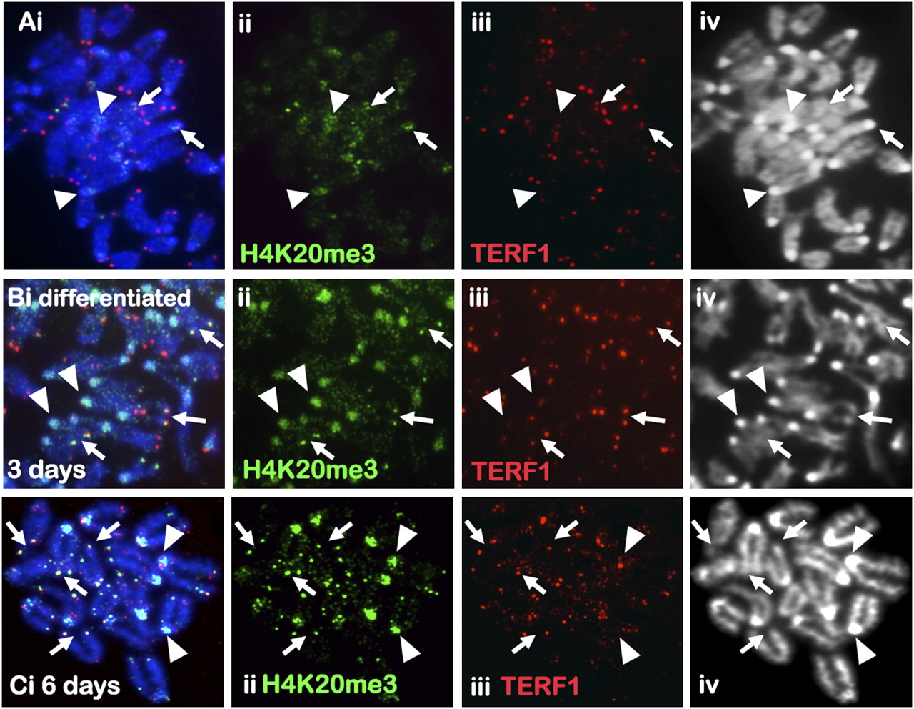
Figure 1.
Immunofluorescence analysis of H4K20me3 in mouse ES cells. (A) H4K20me3 (ii, green; Abcam) staining in ES129.1 cells was weak, with enrichment on pericentric heterochromatin (arrowheads) and on some telomeres (arrows) as indicated by TERF1 staining (iii, red). (B–C) Three days (B) to 6 d (C) of differentiation increased H4K20me3 staining (ii, green) at the pericentric heterochromatin (arrowheads) and telomeres (arrows) in ES129.1 cells.