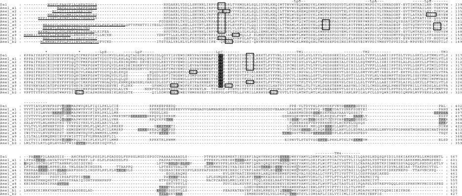
Figure 1.
Protein sequence alignment of A. mellifera nAChR subunits. Dα1 of D. melanagaster is included for comparison. N-terminal signal leader peptides are underlined, and the loops implicated in ligand binding (LpA-F), as well as the four transmembrane regions (TM1–TM4) are indicated. The two cysteines forming the cys-loop are marked by asterisks and the vicinal cysteines characteristic of a subunits are highlighted in black shading. Putative N-glycosylation sites are boxed, potential cAMP, PKC, and CK2 phosphorylation sites are boxed with gray shading, and potential tyrosine kinase phosphorylation sites are enclosed in the gray-shaded ovals. Note that the Amelα3L, Amelα4 exon 4, and Amelα6 exon 8a splice variants are shown in the alignment.