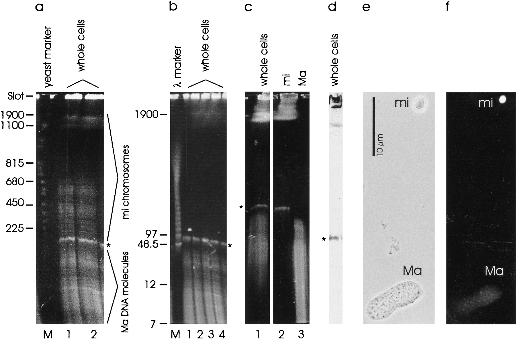
Visualization of Stylonychia DNA by PFGE and nuclear localization of the 90-kb DNA molecule by hybridization analyses. (a,b) DNA from whole Stylonychia cells was separated by PFGE and stained with ethidium bromide (a, lanes1 and 2, and b, lanes1–4). Two different DNA preparations were used ina and b, depending on the DNA preparation and the gel run, to show that the micronuclear chromosomes (mi) are more or less separated, whereas the 90-kb DNA molecule and the macronuclear DNA (Ma), with a size up to 40 kb, are visible on every gel. (c) DNA from whole cells (lane 1) and from isolated micronuclei (lane 2) or macronuclei (lane 3), respectively, was separated by PFGE and stained with ethidium bromide. (d) DNA from whole cells was separated by PFGE, transfered on a nylon filter, and hybridized with labeled micronuclear DNA. (e,f) In situ hybridization of micronuclear DNA with fixed Stylonychia cells. (e) Phase-contrast (bar, 10 μm); (f) in situ hybridization (FITC-labeled antibody against DIG-labeled micronuclear DNA). Sizes are given in kb. Molecular size markers were used: Saccharomyces cerevisiae chromosomes (lane M ina, Pharmacia), 1-kb ladder (7 and 12 kb in b, GIBCO BRL), λ concatemers (lane M in c, Bio-Rad). (*) 90-kb fraction.