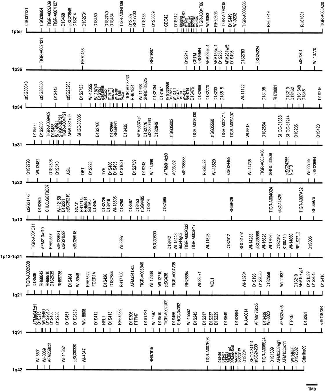
Figure 1.
Chromosome 1 RH framework. Framework markers are listed horizontally from top left to bottom right starting at the 1p terminus. Markers are spaced proportionally to their centiRay positions. Cytolocations are indicated at the beginning of each line. An approximate physical scale is represented at bottom right.