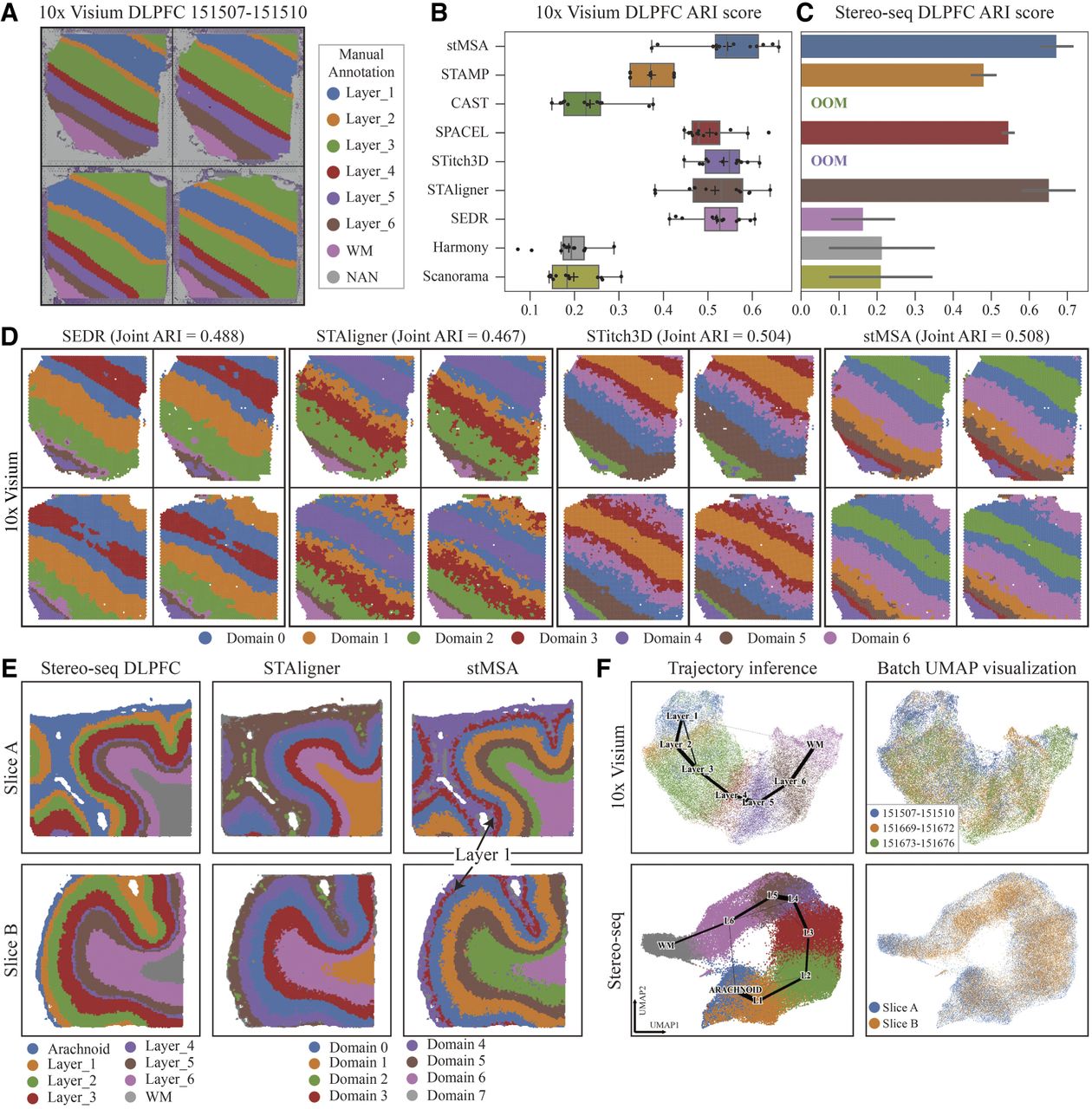
stMSA improves joint domain identification for human dorsolateral prefrontal cortex (DLPFC) slices. (A) Manual annotations, ranging from the cortex layer 1 to the white matter, are provided with the histology image of a donor (slice identifiers: 151,507–151,510). (B) The box plot illustrates the ARI scores across 12 10x Visium DLPFC slices, generated by training separately for each donor. (C) The bar plot illustrates the average ARI scores for the two Stereo-seq DLPFC slices. Error bars represent the 95% confidence intervals of the ARI scores. Notably, STitch3D and CAST were unable to produce results for the Stereo-seq DLPFC data set on our server with 24 GB GPU memory and 128 GB RAM. (D) Visualization of domain detection results for SEDR, STAligner, Stitch3D, and stMSA. The domain detection processes are conducted using all four slices as inputs. (E) The ground-truth, STAligner, and stMSA domain detection results of the two Stereo-seq-obtained DLPFC slices. (F) The trajectory inference plot generated based on the embeddings learned by stMSA using all 12 DLPFC slices for the 10x Visium and the two Stereo-seq DLPFC slices (the color reflects the ground-truth domains). And the UMAP visualization for different donors/slices (the color reflects three donors/slices).