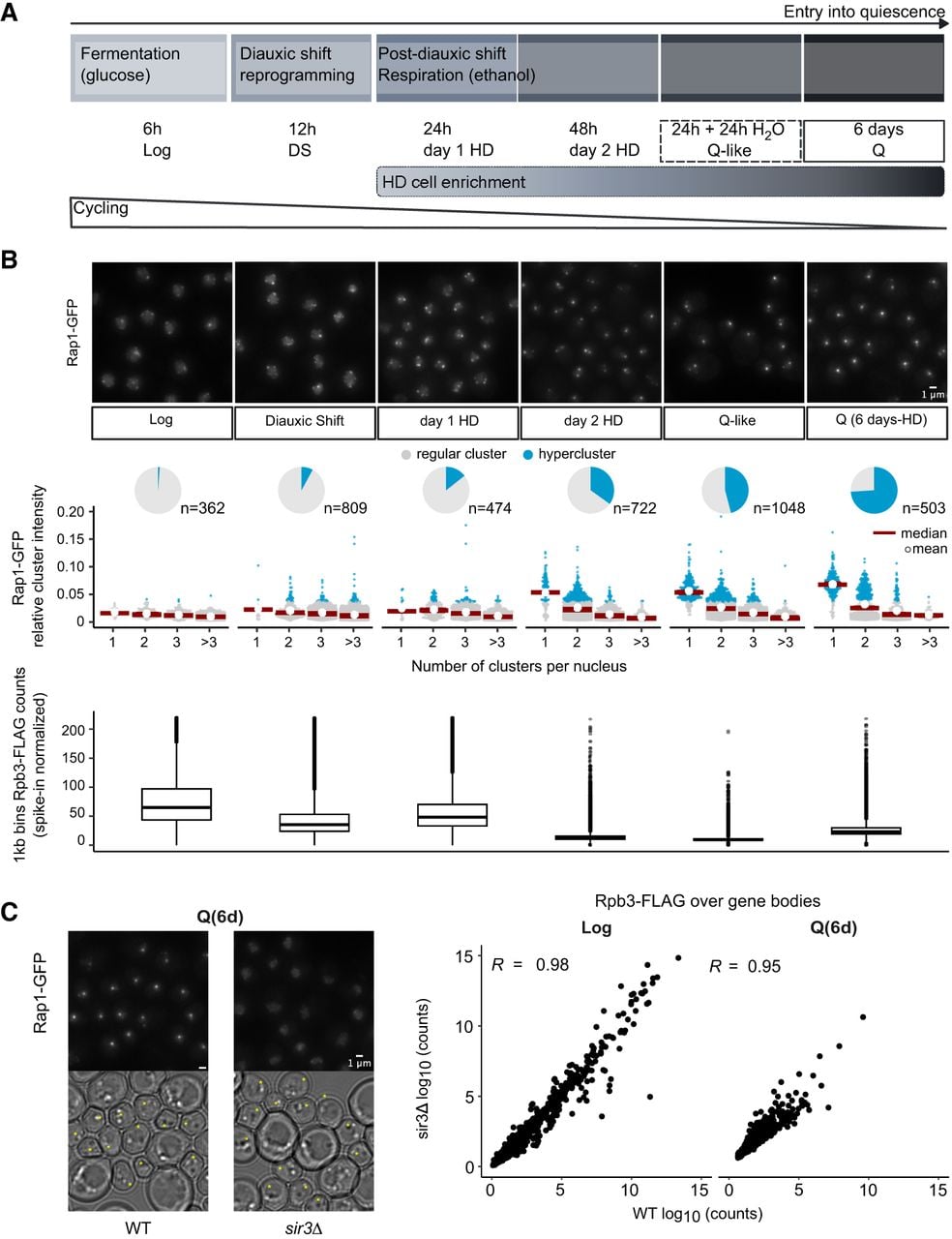
Transcriptional shutdown during quiescence entry occurs independently of telomere hypercluster formation. (A) Timeline of events occurring during quiescence entry. Samples in the nutrient exhaustion kinetics are shown as a function of time after inoculation in rich liquid medium (YPD). All cells following diauxic shift (DS) are sorted through a density gradient (Allen et al. 2006) to recover the highly dense (HD) fraction. For abrupt starvation, yielding Q-like cells, gradient-enriched cells grown for +24 h to post-DS (day1-HD cells) are resuspended in water for 24 h. (B) Proportion of nuclei with hyperclusters during quiescence entry compared with spike-in calibrated RNAPII ChIP-seq (Rpb3-FLAG) counts genome-wide. (Top) Representative images of Rap1-GFP sampled during quiescence entry. Bar = 1 µm. (Middle) Rap1-GFP cluster intensities relative to total nuclear signal classified according to the number of clusters found per nucleus. Hyperclusters, shown in blue, are defined as clusters with a relative intensity more than fourfold higher than the average cluster in Log phase. Pie charts represent the proportion of nuclei with a hypercluster. (Bottom) Rpb3-FLAG spike-in calibrated IP counts averaged over 1 kb bins genome-wide. (C) Comparison of telomere hyperclustering and Pol II (Rpb3-FLAG) binding in quiescence (6d-HD; Q). (Left) Representative images of Rap1-GFP in WT and sir3Δ samples in Q cells. Small cells, with a Feret's diameter <4.5 µm are marked with an asterisk in transmitted light images. Bar = 1 µm. (Right) Rpb3-FLAG counts over genes normalized by gene size in WT versus sir3Δ samples.