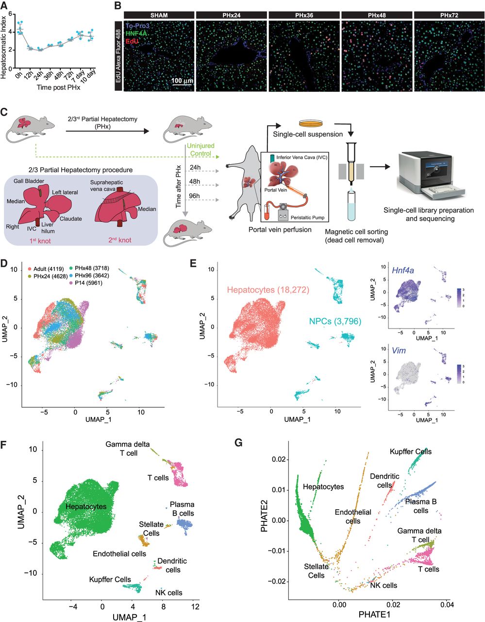
Single-cell analysis of resident hepatic cell populations from immature, adult, and regenerating mouse livers. (A) Time course plot showing the restoration of liver-to-body weight ratio after partial hepatectomy (PHx). The liver recovers its original mass within 7 d after PHx (n = 5). (B) Fluorescent imaging of hepatocyte proliferation measured by in vivo EdU incorporation in post-PHx and Sham livers. White arrows indicate proliferating hepatocytes (colabeled for HNF4A in green, incorporated EdU in red, and nuclei in blue). Images taken under 20× resolution are shown. (C) Overview schematic demonstrating workflow for isolation of mouse liver cells for single-cell RNA sequencing (scRNA-seq). Portal vein perfusion of collagenase containing buffer was used to isolate single liver cells from uninjured P14 pups and adults as well as mice at 24, 48, and 96 h after 2/3rd PHx (n = 1/time point). Single-cell library preparation was performed with whole-cell suspensions individually for each mouse using a 10x Genomics Chromium Single Cell 3′ Reagent Kit (V3 chemistry) after magnetic-activated cell sorting to remove dead cells. The inset details our PHx procedure, showing the position of two knots before excision of the respective liver lobes. (D) Combined UMAP projection of all 22,068 cells identified after QC cutoffs and batch correction. Cells are colored by the batch of origin, and the total number of cells identified from each batch is given in parentheses. (E) Identification of hepatocyte and nonparenchymal cell (NPC) subpopulations. Graph-based clustering in Seurat v3.1 followed by marker gene analysis revealed broad epithelial and nonepithelial cellular identities. Feature plots shown as insets show higher expression of expression of Hnf4a (a hepatocyte marker) and Vim (a nonepithelial marker) especially in populations identified as hepatocytes and NPCs, respectively. (F) Combined UMAP projection of all cells, colored by the annotated cell type. (G) PHATE projection of the ∼22,000 cells from different stages of liver development and regeneration. Cells are colored by annotated cell types from F.