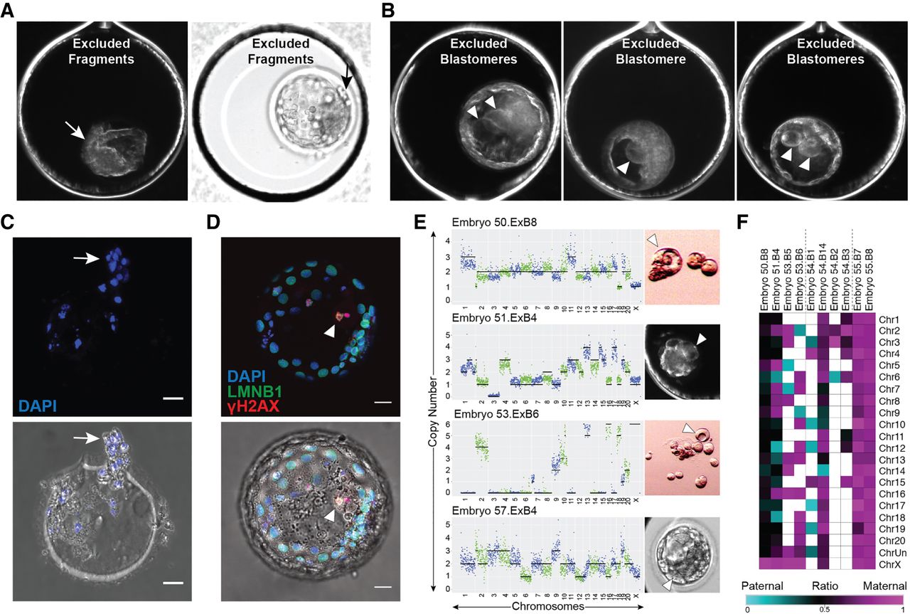
Cellular fragments and aneuploid blastomeres are excluded upon blastocyst formation. Time-lapse image frames from two rhesus blastocysts showing exclusion of several cellular fragments to the perivitelline space of the embryo (A; arrow) or of three rhesus blastocysts with one to two nondividing excluded blastomeres in the blastocoel cavity (B; arrowheads). (C) The zona pellucida of the blastocyst that showed cellular fragment exclusion with remaining DNA positive for DAPI (blue) staining following hatching (white arrow). (D) A blastocyst with blastomere exclusion immunostained for LMNB1 (green) and γH2A.X (red) using DAPI as a marker for DNA. The large excluded blastomere appeared binucleated with strong γH2A.X signals (white arrowhead), indicating that double-stranded DNA breaks had occurred. Brightfield image with immunofluorescence overlay provided below. (E) Additional examples of large excluded blastomeres (right; white arrowheads) collected during the morula-to-blastocyst transition for sequencing. CNV analysis (left) determined that each excluded blastomere had chaotic aneuploidy. Scale bars, 25 µm. (F) Heat map of maternal versus paternal SNP allele ratios in excluded blastomeres shows parental origins.