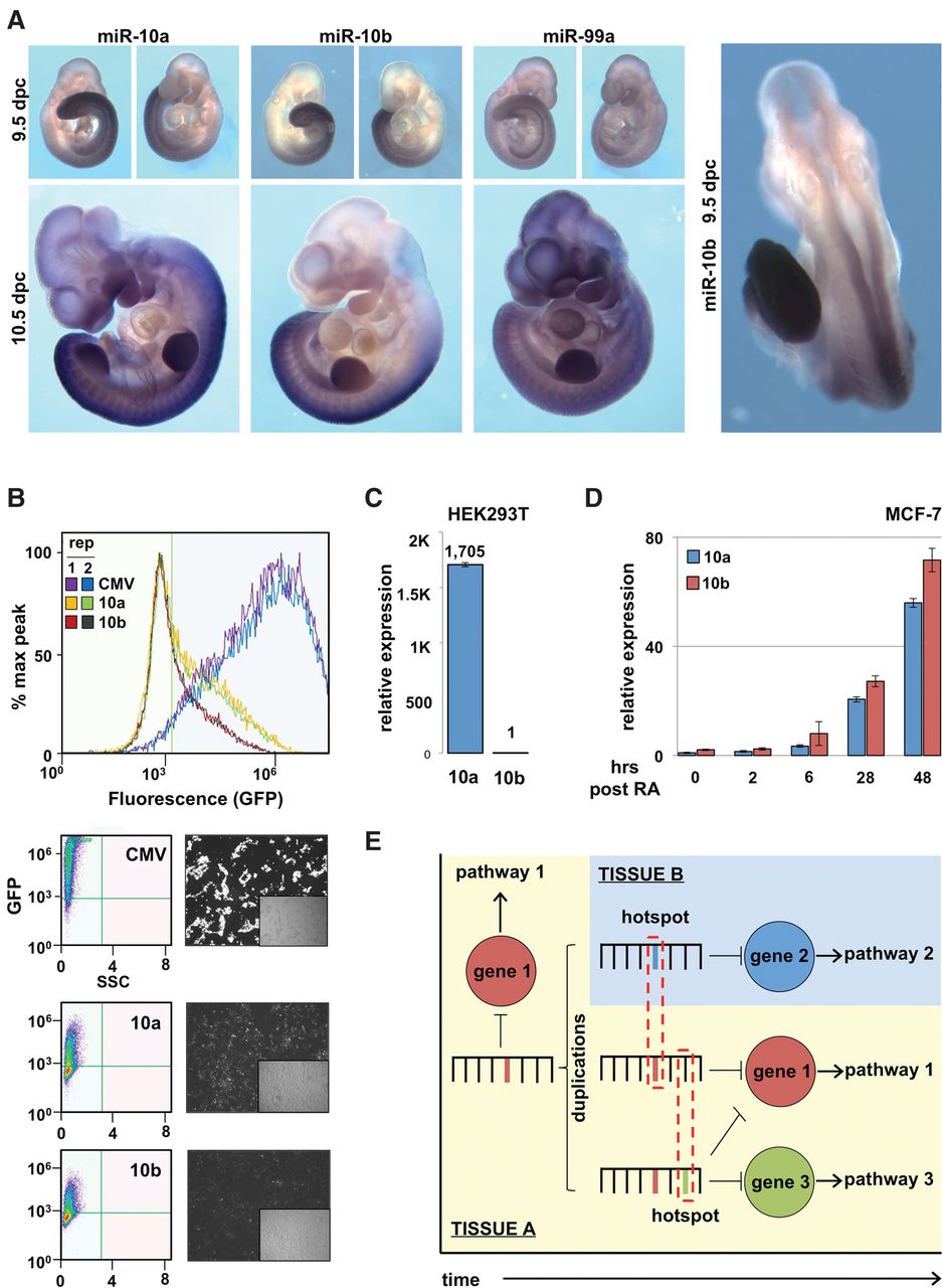
miR-10 family members have divergent expression patterns. (A) Whole-mount in situ hybridizations of mouse embryos at 9.5 and 10.5 d post-coitus (dpc), using LNA-based probes to detect the expression pattern of mature miR-10a, miR-10b, and miR-99a. Data are representative of three embryos. (B) We cloned the promoter regions (2000 bp) of MIR10A (10a) and MIR10B (10b) and tested their ability to drive GFP expression by transient transfection in HEK293T cells. CMV promoter was used as a positive control to indicate transfection efficiency. Fluorescence quantified by flow cytometry (n = 2) and representative fluorescent images of cells. (C) qRT-PCR of mature miR-10a (10a) and miR-10b (10b) levels in HEK293T cells (n = 3). (D) Expression dynamics of mature miR-10a (10a) and miR-10b (10b) following 10 μM retinoic acid in MCF-7 cells (n = 3). (E) Model: Over evolutionary time, miRNAs evolve to regulate specific mRNAs within pathways. Following a miRNA duplication event, there is frequent sequence divergence between paralogous miRNAs, along with the emergence of novel miRNA expression patterns. The sequence of target genes expressed in these novel contexts may influence specific nucleotide changes to facilitate more efficient repression by the duplicated miRNA.