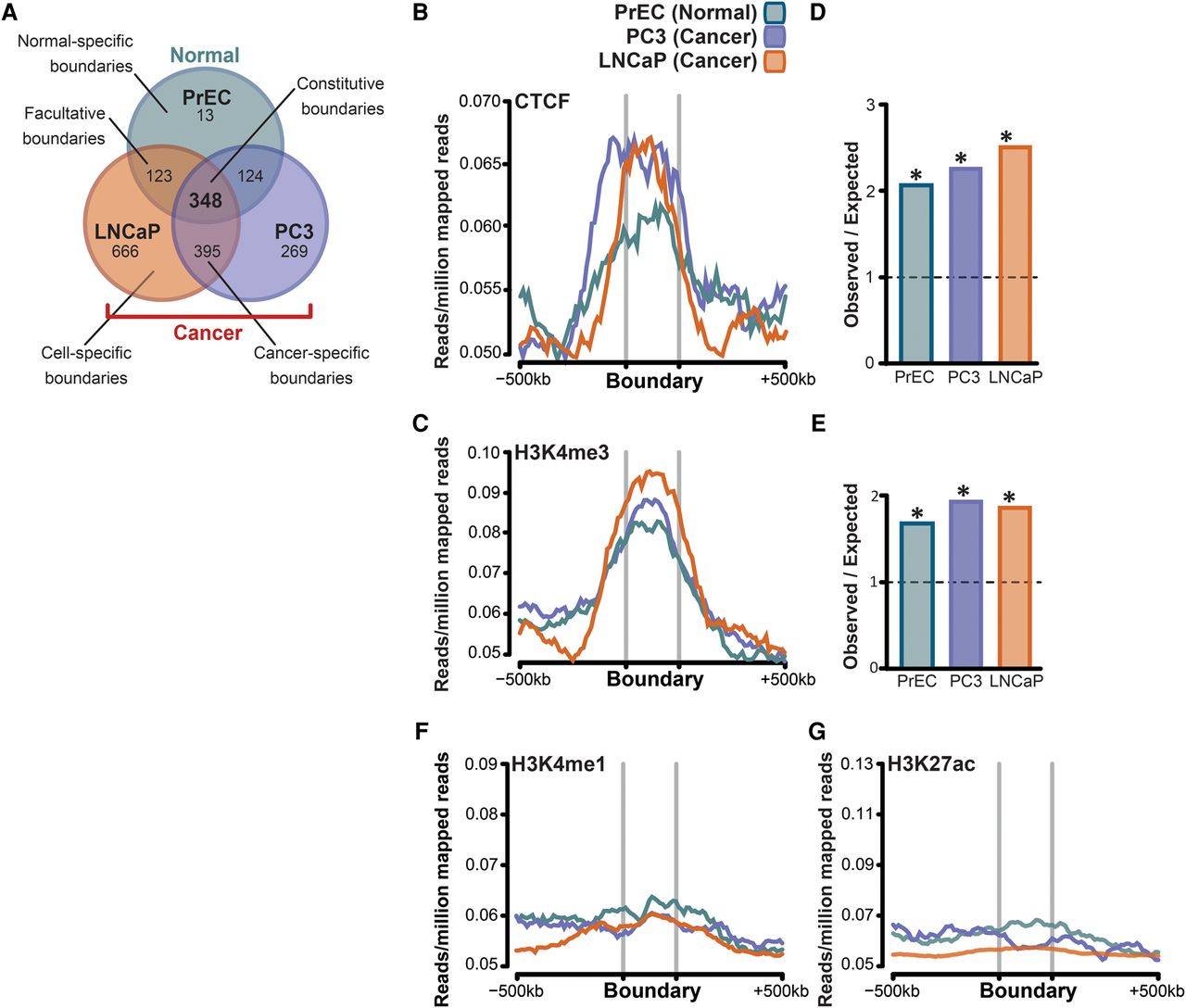
Cancer cells acquire unique topological domain boundaries that retain CTCF binding and enrichment for H3K4me3. (A) Venn diagram showing that the majority of domain boundaries that are present in normal cells (PrEC) are also present in cancer cells (constitutive boundaries; PC3 and LNCaP), while ∼20% of boundaries were maintained in only one of the two cancer cells (cell-type–specific boundaries). (B,C,F,G) Genome-wide average distribution of CTCF, H3K4me3, H3K4me1, and H3K27ac binding around the domain boundaries in PrEC, PC3, and LNCaP cells. (D) Fold enrichment of CTCF binding at the domain boundaries in PrEC, PC3, and LNCaP cells compared to that expected by chance. (E) Fold enrichment of H3K4me3 binding at the domain boundaries in PrEC, PC3, and LNCaP cells compared to that expected by chance.