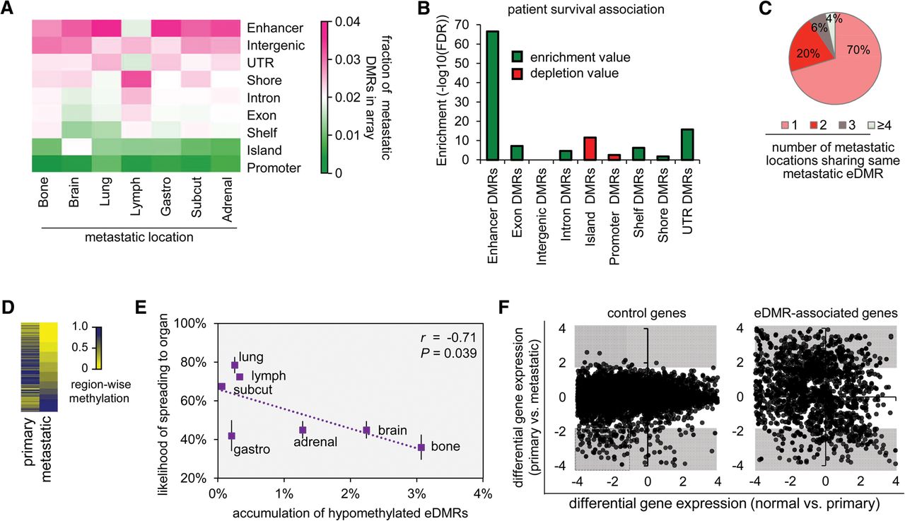
Accumulation of eDMR hypomethylation correlates with likelihood of metastasis and patient outcome. (A) Heat map shows that enhancers are more differentially methylated than other genomic features (y-axis; sorted according to fraction of DMRs in each category) (see Fig. 1A). (Adrenal) Metastases to adrenal glands, (gastro) metastases to the gastrointestinal tract, (lymph) metastases to lymph nodes, (subcut) subcutaneous metastases. (B) Enhancers are significantly more enriched with DMRs that can differentiate between patient survival times (see Methods). Green and red bars represent enrichment and depletion, respectively. Y-axis represents the −log10(FDR-corrected P-values) significance (binomial distribution). (C) Most melanoma eDMRs (70%) are exclusive to one metastatic site (see Fig. 2A). (D) Heat map shows that most eDMRs are hypomethylated between primary and metastatic melanoma (74%). (E) Accumulation of eDMR hypomethylation is negatively correlated with the likelihood of forming melanoma metastases at each body location (Pearson's correlation, r = −0.71, P = 0.039, one-sided hypothesis testing). (F) Compared to all other genes (control genes; y-axis, left panel), genes associated with eDMRs (y-axis, right panel) are differentially expressed between primary melanoma and metastatic melanoma (57% and 3%, respectively; marked by gray areas), whereas, differential expression between melanocytes and primary melanoma is similar for both groups (control genes, 53%, left panel; eDMR-associated genes, 60%, right panel; x-axes). All parts of the figure refer to differential methylation between patients with primary melanoma and patients with metastatic melanoma.