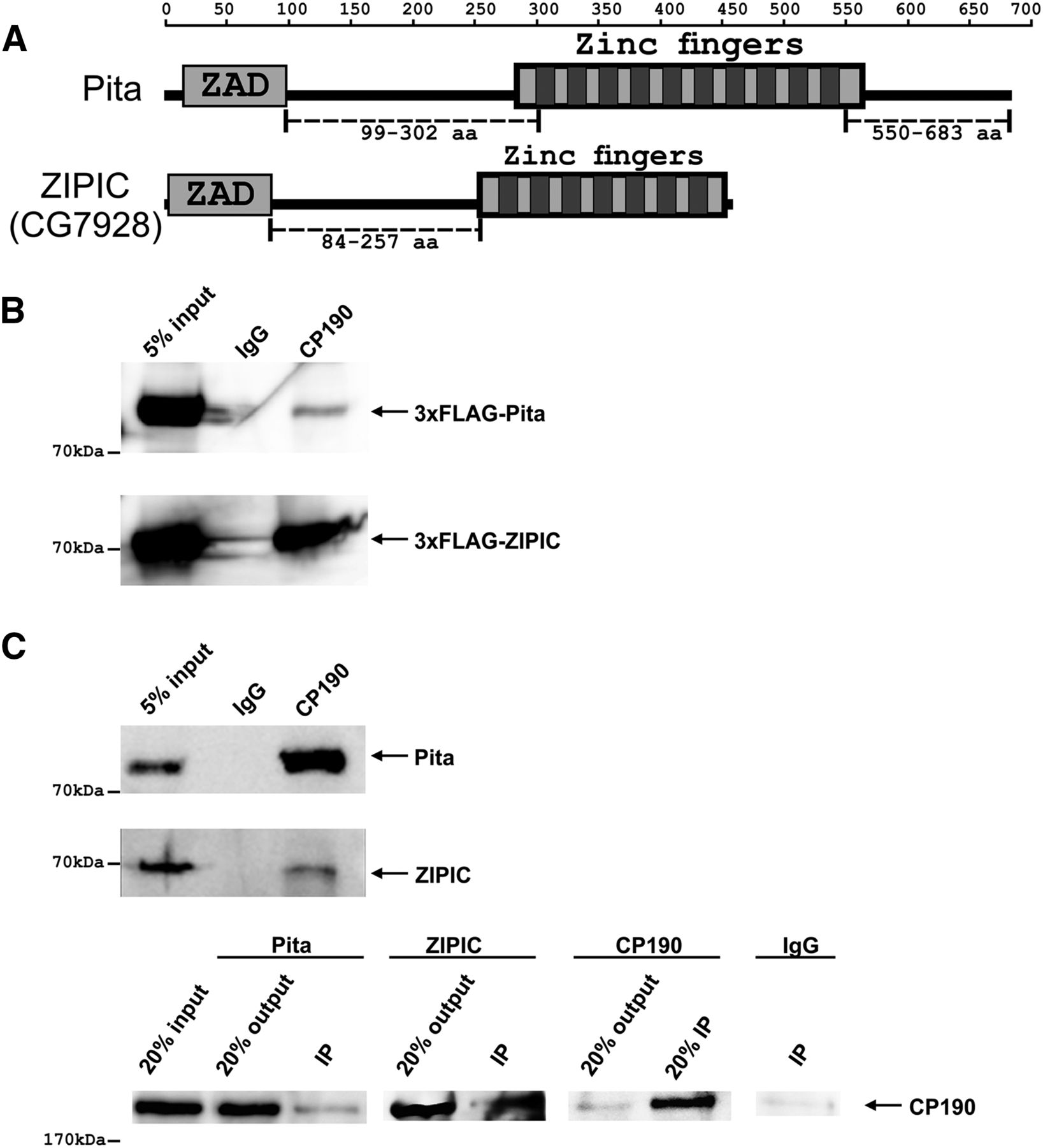
Interaction of CP190 with Pita and ZIPIC proteins. (A) Structure of full-length Pita and ZIPIC proteins containing the ZAD domain and C2H2-type zinc fingers. The scale shows the number of amino acid residues. Broken lines indicate regions used to prepare antibodies. (B) Nuclear extracts from Drosophila S2 cells cotransfected with CP190 and 3 × FLAG-Pita/ ZIPIC were immunoprecipitated with antibodies against CP190 (using nonspecific IgG as a negative control), and the immunoprecipitates were analyzed by Western blotting for the presence of FLAG-tagged proteins. (C) Nuclear extracts from Drosophila embryos were immunoprecipitated with antibodies against CP190, Pita, or ZIPIC (using nonspecific IgG as a negative control), and the immunoprecipitates (IP) were analyzed by Western blotting for the presence of Pita, ZIPIC, and CP190. Inputs show the starting samples of nuclear extract; outputs are supernatant after sedimentation of immunoprecipitated material.