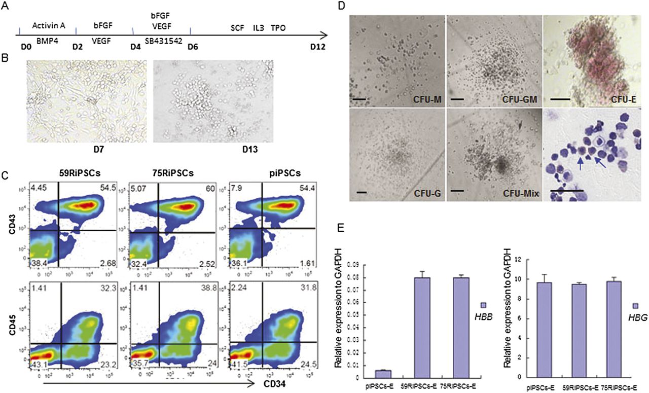
Hematopoietic differentiation of gene-corrected iPSCs. (A) Schematic representation of a stepwise hematopoietic differentiation strategy for iPSCs. (B) Representative morphology changes at day 7 and day 13 in hematopoietic differentiation of corrected iPSC lines. (C) Flow cytometric analysis of piPSCs (parental lines), 75RiPSCs, and 59RiPSCs lines harvested at day 14 showing the specific hematopoietic stem and progenitor markers: CD34+, CD43+, and CD45+. (D) Colony-forming assay for differentiated cells at day 10 revealed various types of hematopoietic colonies as well as nucleated cells, including erythroblasts. Blue arrows indicate erythroid cells. (E) HBB and HBG gene expression (normalized to GAPDH) were measured by quantitative RT-PCR in hematopoietic differentiation of parental lines (piPSCs-E), −28 mutation-corrected (59RiPSCs) lines, and 41/42 mutation-corrected (75RiPSCs) lines (data represent mean ± SEM, n = 3).