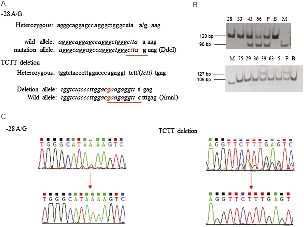
Figure 3.
Identification of gene correction in β-thalassemia iPSCs. (A) Nucleotides changed (in red) in PCR primers to generate restriction enzyme sites to distinguish the normal from the mutant alleles. For the –28 location, replacing nucleotide “a” with “t” at –31 generates a DdeI site for the mutant allele. At 41/42, replacing “cc” with “ga” generates an XmnI site for the wild-type allele. (B) PCR amplification followed by restriction enzyme digestion reveals a normal sequence for clones 28 and 33 at the –28 site and clones 75, 29, 36, 63, and 5 at the 41/42 location. (P) Parental iPSCs, (B) BAC, (M) marker. (C) Sequences of the two mutation sites showing correction of the heterozygous states (upper) to the normal sequences (lower).