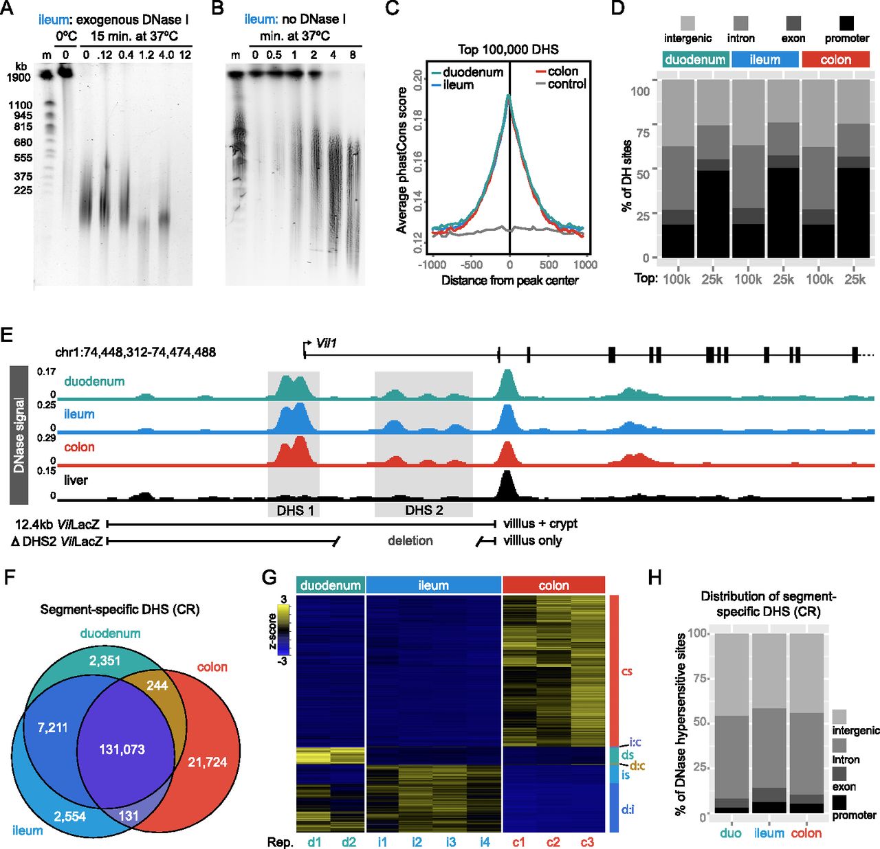
Endogenous DNase activity distinguishes open chromatin in mouse intestinal epithelial cells. (A) Pulse-field gel image of nuclei digested for 15 min at 37°C with increasing concentrations of exogenous DNase I. Note that high-molecular-weight (HMW) DNA is stable at 0°C; however, there is significant DNA digestion even with no addition of exogenous DNase when nuclei are incubated for 15 min at 37°C. (m) Yeast chromosome marker. (B) Endogenous DNase activity is detected within 30 sec after moving nuclei to 37°C, and by 8 min, most HMW DNA is digested. Patterns were consistent for duodenum, ileum, and colon (see Supplemental Fig. S4). The observed digestion pattern is similar to reported digestion patterns using exogenous DNase I (Song and Crawford 2010). For DNase-seq library preparation, nuclei digested for 2, 4, and 8 min were pooled to capture a range of DNase hypersensitivities. Libraries were prepared for duodenal, ileal, and colonic IECs. (C) Average phastCons scores plotted for the top 100,000 DHSs from duodenal, ileal, and colonic IECs centered at the peak maximum. Nongenic DNA flanking ileal DNase hypersensitive sites (DHSs) was used to assess background conservation (control). (D) Feature distribution of the top 100,000 and 25,000 DHSs from each tissue. Note the increased representation of promoter-associated sites (<2 kb from annotated transcription start sites) in the 25,000 DHSs with the highest signal intensity. (E) DNase-seq signal tracks from conventionally raised (CR) duodenal, ileal, and colonic IECs at the villin 1 (Vil1) locus. Note strong peaks at the transcription start site (DHS 1) and within the first intron (DHS 2). A 12.4-kb region including both DHS 1 and DHS 2 drives IEC-specific crypt and villous expression in the duodenum, ileum, and colon (Madison 2002); however, DHS 2 is required for crypt expression. For comparison, DNase-seq signal from the liver is also shown. (F) Venn diagram enumerating differential DHSs along the length of the GI tract. (G) Hierarchical clustering of differential DHSs across replicates of CR duodenal, ileal, and colonic IECs reveals open chromatin sites specific to each tissue. (cs) Colon-specific; (i:c) ileum and colon; (ds) duodenum specific; (d:c) duodenum and colon; (is) ileum specific; (d:i) duodenum and ileum. (H) Feature distribution showing that the majority of segment-specific DHSs are located in intergenic (>2 kb away from a gene body) or intronic regions of the genome. See also Supplemental Figures S2–S4 and Supplemental Tables S4 and S6.