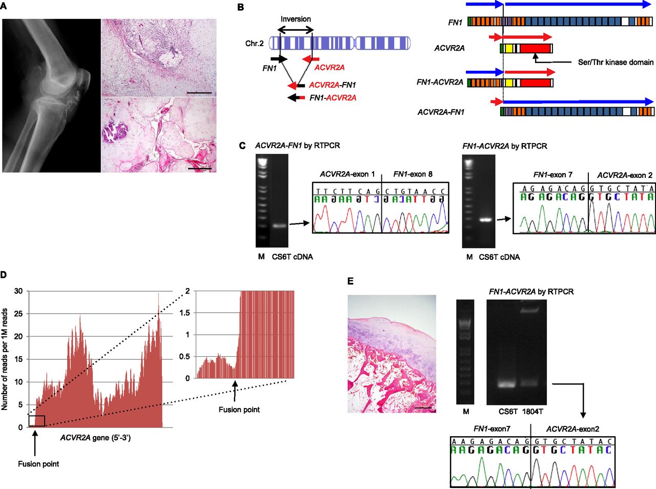
FN1-ACVR2A fusion gene in chondrosarcoma. (A) Synovial chondrosarcoma case. (Left panel) Radiograph revealed a calcified soft tissue mass surrounding the knee joint without bone involvement. (Right, lower panel) This hypercellular myxoid tumor diffusely infiltrated periarticular and subcutaneous tissues, dissecting through fat and collagen (scale bar, 500 μm). (Right, upper panel) The tumor focally resembled synovial chondromatosis (scale bar, 1 mm) and later metastasized to the inguinal lymph node. (B) Schematic presentation of an intrachromosomal inversion in chromosome 2q (left) and two in-frame fusion proteins generated by this rearrangement. (C) Validation of fusion transcripts (left, ACVR2A-FN1, and right, FN1-ACVR2A) by RT-PCR and Sanger sequencing. (D) Estimation of ACVR2A gene expression at nucleotide resolution by counting RNA sequencing reads. Note the sharp increase in AVCR2A expression after the fusion point. (E, left) Histology of the multiple osteochondroma case. This tibial tumor is composed of exophytic bony growth capped by benign hyaline cartilage (scale bar, 1 mm). (Right) Validation of the FN1-ACVR2A fusion transcript by RT-PCR and Sanger sequencing in an osteochondromatosis case (1804T).