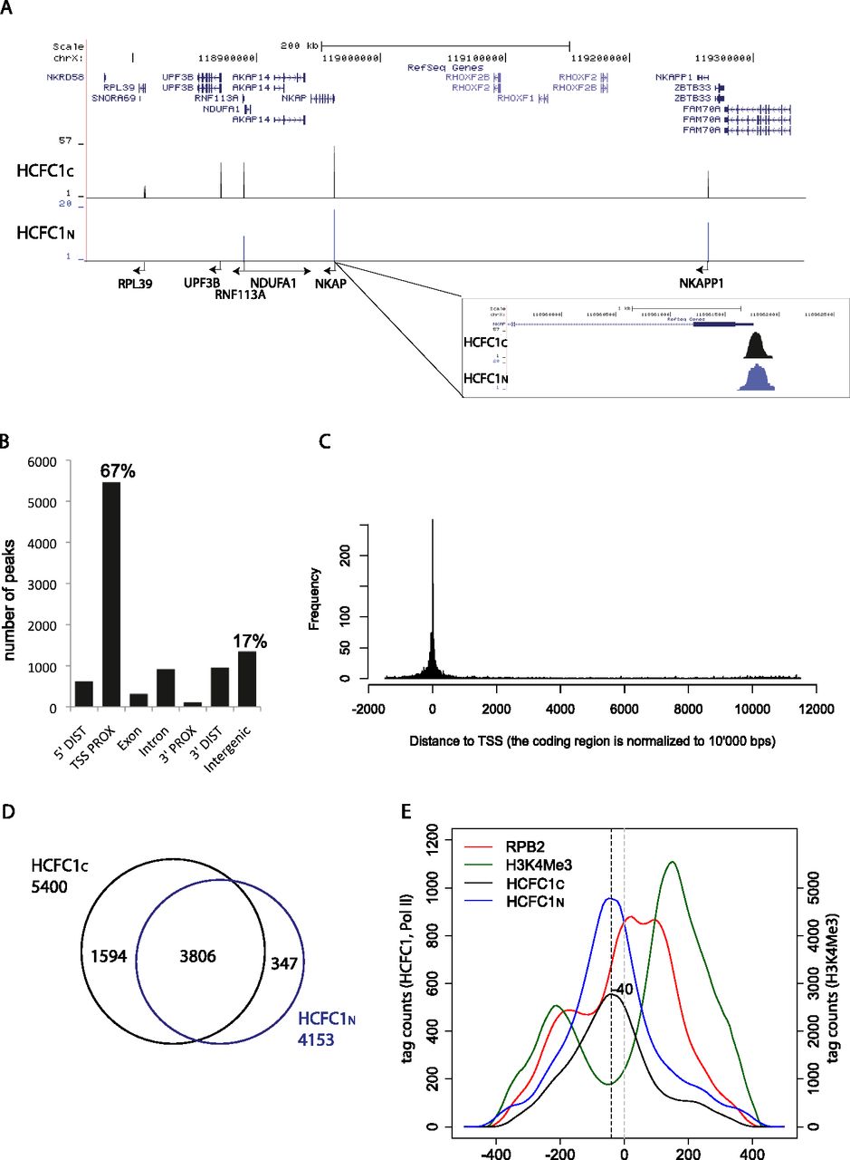
Genomic localization of human HCFC1. (A) Typical profile of HCFC1 peaks throughout the genome (UCSC Genome Browser) and magnification of one HCFC1 peak. Both HCFC1C and HCFC1N data are shown. (B) Distribution of the HCFC1C peaks according to their genomic localization. (5′ DIST) −5000 to −1000 of TSSs; (TSS-PROX) −1000 to +500 of TSSs; (3′ PROX) −500 to +1000 of end of transcript; (3′ DIST) +1000 to +5000 of end of transcript; exon and intron within the coding sequence only; (Intergenic) >5000 bp distal of annotated transcript regions. Percentages of the total number of peaks are indicated for the TSS-PROX and intergenic regions. (C) Enrichment of HCFC1 peaks within and surrounding annotated transcript regions. All transcribed regions are normalized to 10,000 bp. (D) Venn diagram showing the number of shared and distinct RefSeq TSSs bound by either HCFC1C or HCFC1N. (E) Cumulative mapping of HCFC1N (blue), HCFC1C (black), Pol II (RPB2; red), and H3K4Me3 (green) binding sites within −250 to +250 of TSSs. Binding sites were mapped as the center of ChIP-seq fragments artificially extended to the experimentally determined average fragment length. (Left axis) Corresponds to HCFC1N, HCFC1C, and Pol II distribution; (right axis) corresponds to H3K4Me3 distribution. The most enriched position for HCFC1C is indicated as the base pair distance from the TSS and by the dashed black vertical line. TSS is indicated by the dashed gray vertical line.