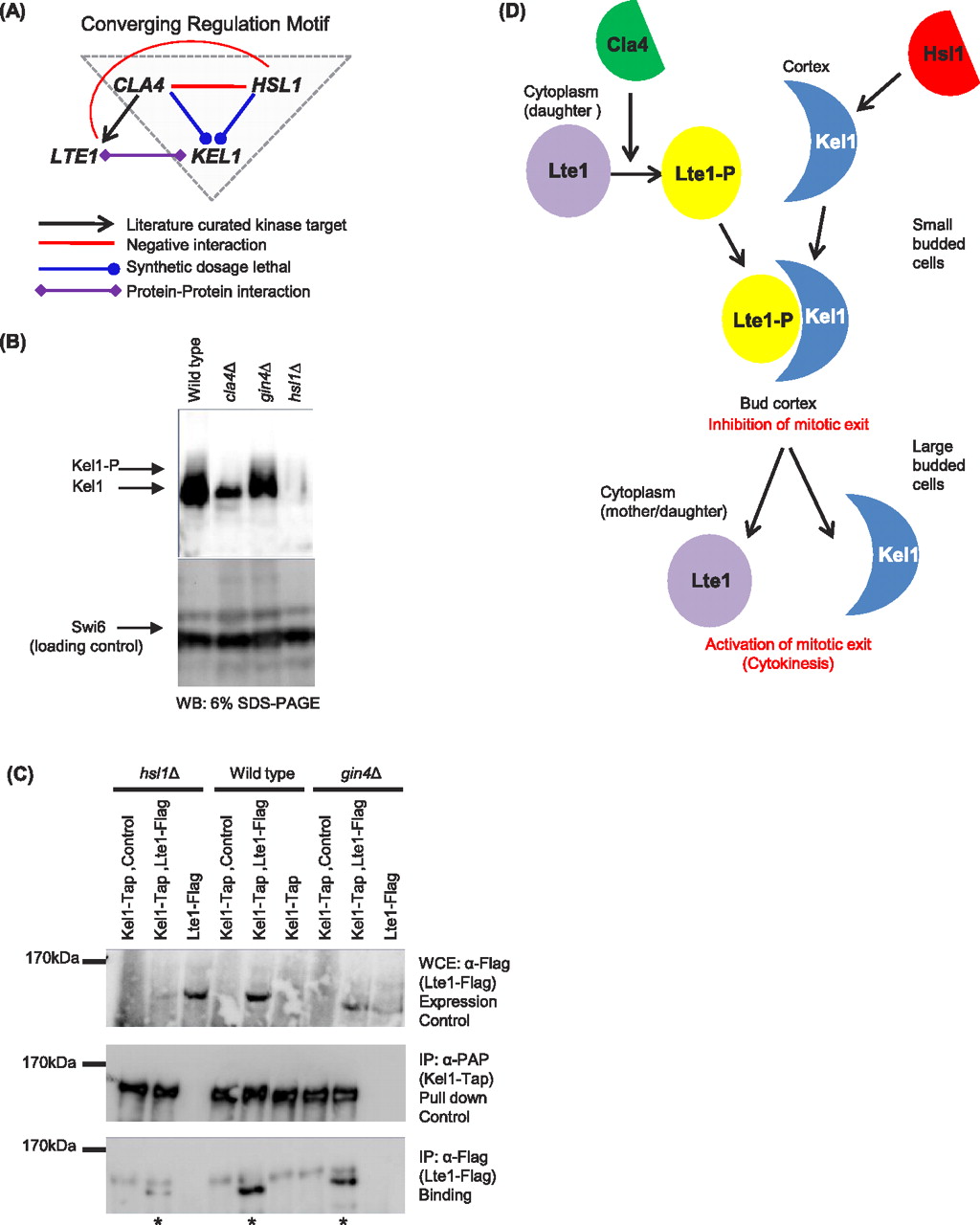
Kel1 protein is co-regulated by budneck kinases. (A) Overrepresented motif (gray triangle), supporting a functional connection between Kel1 and the two budneck kinases Cla4 and Hsl1 in a converging regulation model. (B) Endogenous Kel1 protein levels are reduced in hsl1Δ and cla4Δ but not in gin4Δ mutant strains. Kel1-TAP protein was immunoprecipitated from the indicated budneck kinase mutant strains and Western blots were probed with anti-PAP to detect total Kel1 protein. Electrophoretic mobility shift of Kel1 protein is shown (Kel1-P). Swi6 was used as a loading control. (C) Integrated Kel1-TAP protein was immunoprecipitated in the presence of Lte1-Flag and association of Lte1 with Kel1 was detected using α-flag antibody. The amounts of Lte1-Flag in the whole cell extract (WCE; top panel) and in the Kel1 immunoprecipitate (bottom panel) are shown. (Middle panel) The amount of Kel1-TAP in the immunoprecipitate (anti-PAP). Immunoprecipitation assays were performed in wild-type, hsl1Δ, gin4Δ background strains. cla4Δ strain was not tested, due to severe toxicity upon Kel1-TAP expression. (D) Model for regulation of Kel1 by budneck kinases. Lte1 is phosphorylated by Cla4, which triggers binding to its anchor, Kel1. Hsl1 regulates the stability of Kel1 protein, which allows the anchor to become available for binding to Lte1-P. Together, the complex inhibits mitotic exit possibly to delay budding to allow the cell to reach the correct size. Later, Cdc14 phosphatase releases Lte1 from its anchor in the cortex into the cytoplasm in order to activate mitotic exit in large budded cells.