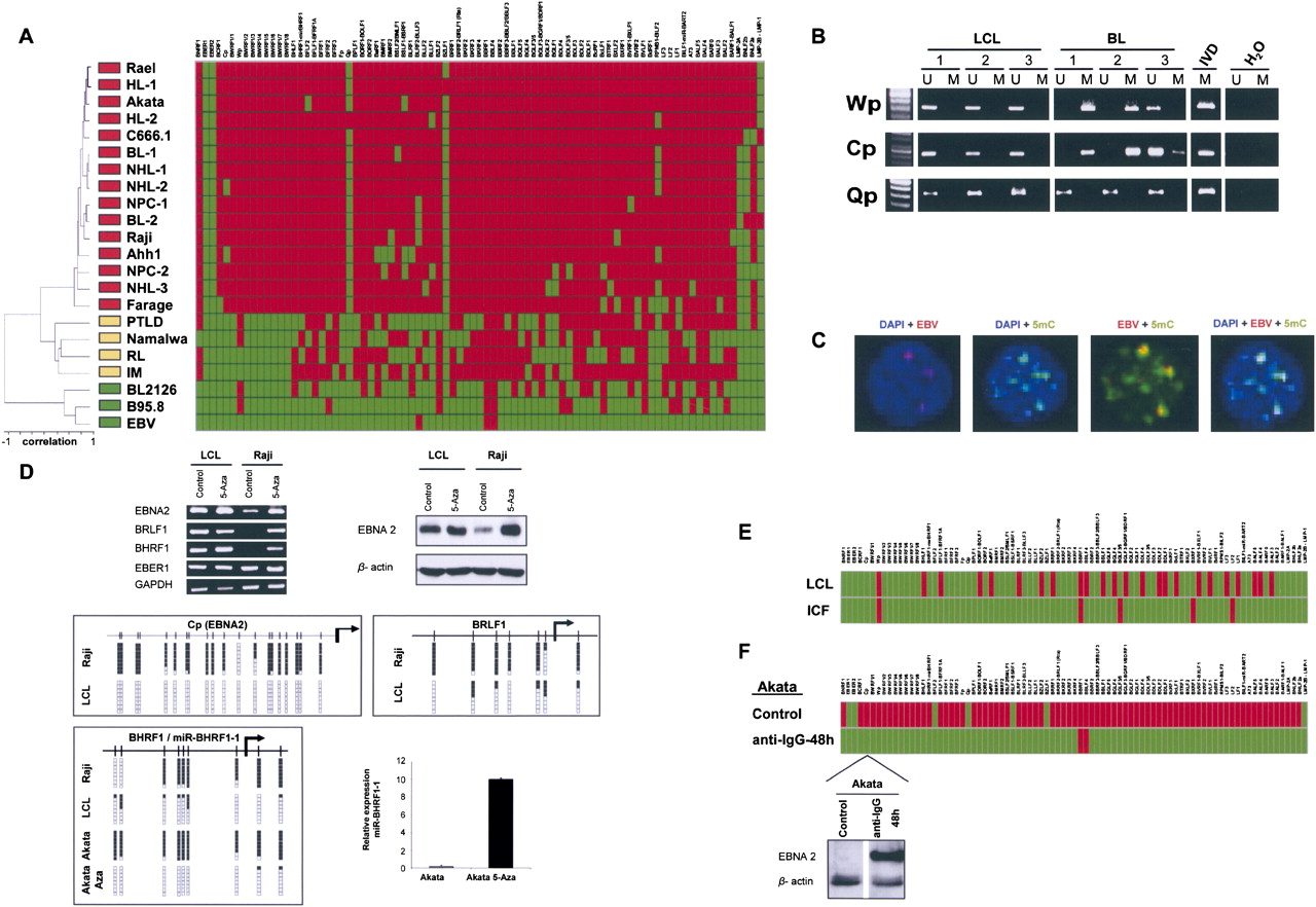
The DNA methylome of EBV. (A) Unsupervised clustering analysis of the EBV DNA methylome in the wild EBV, the B95-8 type EBV, lymphoblastoid cell line (BL2126), benign lesions ([RL] reactive lymphadenitis in tonsil and [IM] infectious mononucleosis), post-transplant lymphoproliferative disorder (PTLD), primary lymphomas ([NHL] non-Hodgkin lymphoma, [HL] Hodgkin lymphoma, [BL] Burkitt's lymphoma), lymphoma cell lines (Rael, Akata, Raji, Ahh1, Farage, Namalwa), nasopharyngeal primary carcinoma (NPC), and cancer cell line (C666.1). (Red) Methylated and (green) unmethylated CpG dinucleotides encompassing the corresponding 5′-end transcription start sites. (Top) The EBV genome. (B) Methylation-specific PCR analysis of the EBV FP, WP, and CP 5′-end CpG islands in lymphoblastic and Burkitt's lymphoma cell lines. The presence of a band under the U or M lanes indicates unmethylated or methylated sequences. In vitro methylated DNA (IVD) is shown as a positive control. (C) Colocalization of the EBV viral genome determined by FISH and the 5-methylcytosine DNA staining in Akata cells. (D) 5′-CpG island methylation-associated silencing of the EBNA2, BRLF1, and BHRF1-H2H3 genes and the microRNA miR-BHRF1-1 and release of silencing upon the use of a DNA demethylating agent (5-aza-2′-deoxycytidine). (Top) RT-PCRs and Western blots; (middle) bisulfite genomic sequencing; (bottom) q-RT-PCR for miR-BHRF1-1. (E) EBV-immortalized ICF lymphocytes (DNMT3B defective) demonstrate a hypomethylated EBV genome in comparison to the one observed in DNMT3B-proficient EBV-transformed lymphocytes (LCL). (Red) Methylated and (green) unmethylated CpG dinucleotides encompassing the corresponding 5′-end transcription start sites. (F) The induction of the EBV lytic cycle in a lymphoma cell line (Akata) upon addition to the media of anti-IgG caused a massive DNA hypomethylating event in the EBV-transcription start sites. (Red) Methylated and (green) unmethylated CpG dinucleotides encompassing the corresponding 5′-end transcription start sites. (Bottom) Example of Western blot for the EBNA2 protein showing the restoration of expression upon the demethylation of the corresponding CpG island.