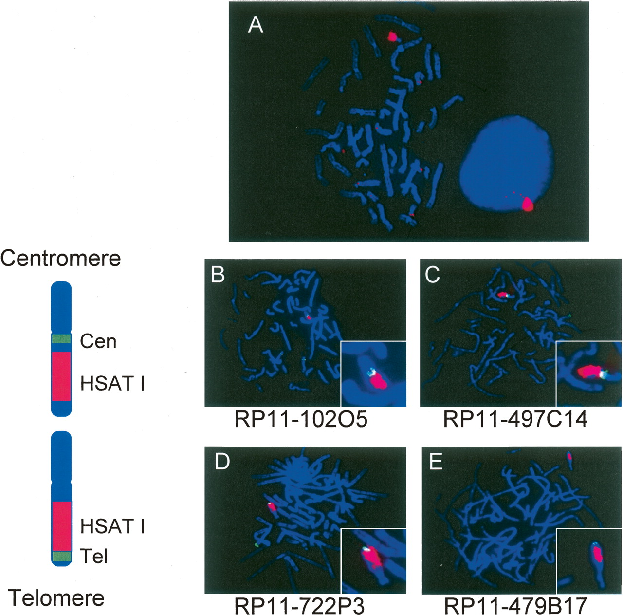
Painting of chromosome Yq12. (A) FISH mapping on DAPI-stained metaphase and interphase human male peripheral blood lymphocytes with the 400-bp cloned PCR product of HSAT I, shown here in red. A signal was detected on chromosome Y. In the interphase cell, the hybridization of the HSAT I probe occurs near the periphery of the nucleus. We had two-dimensional images. Thus, we could not be sure of the position in all the nuclei. We saw the peripheral location in >60%–70% of 100 nuclei examined in separate experiments. (B) Positioning of HSAT I on chromosome Y. FISH mapping on DAPI-stained metaphase human male peripheral lymphocytes showing the position of HSAT I on the Y chromosome. (B,C) The BACs RP11-102O5 and RP11-497C14 (green) map more centromeric on chromosome Y than HSAT I (red). (D,E) The BACs RP11-722P3 and RP11-479B17 (green) map more telomeric on chromosome Y than the HSAT I probe. On the left of the FISH mapping pictures are the ideograms orientating the probes on the chromosome. HSAT I is localized on chromosome Y in the q12 band.