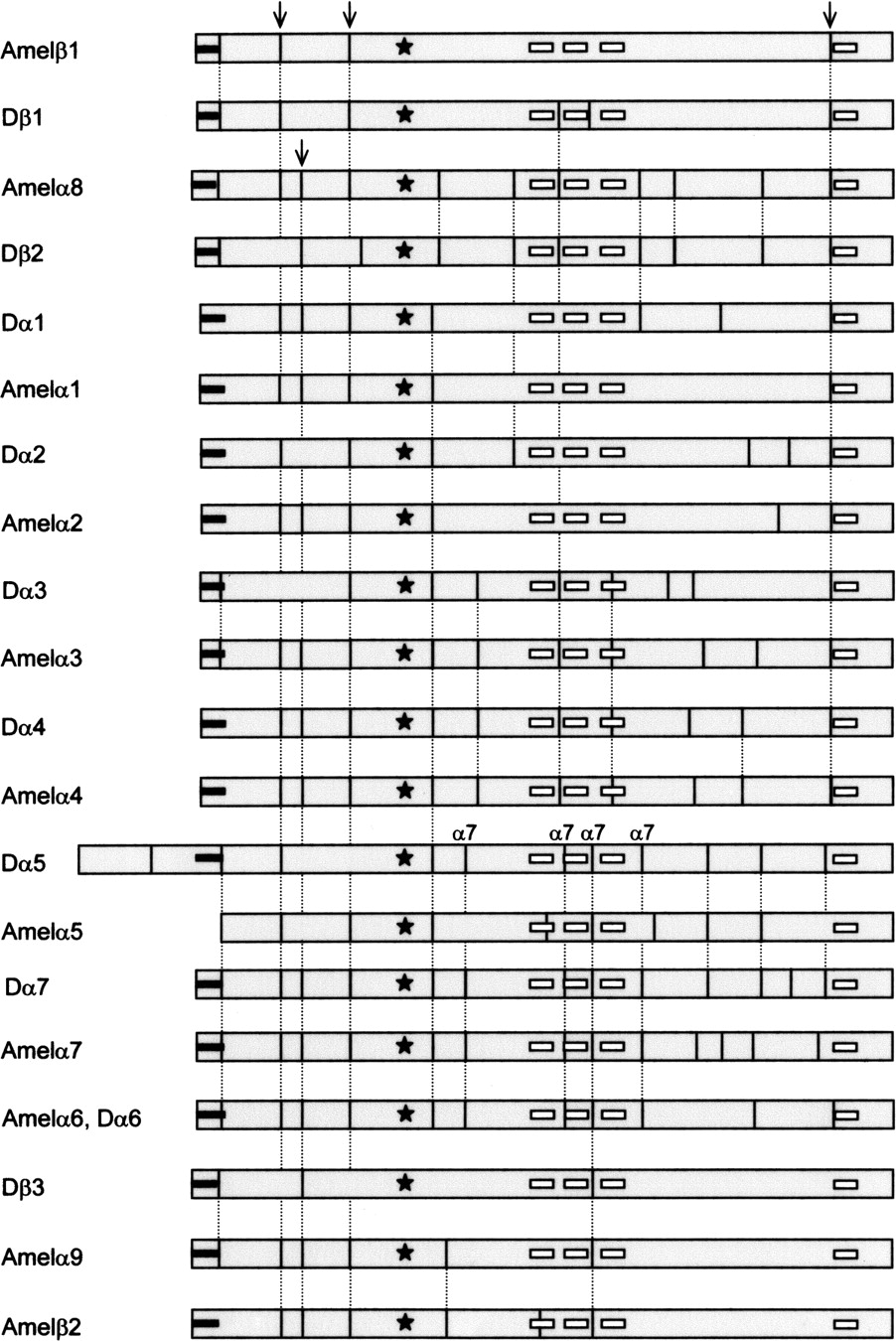
Figure 3.
Exon composition of D. melanogaster and A. mellifera nAChR subunits. The N-terminal signal peptide is shown as a bar, the cys-loop is denoted by a star, and the four transmembrane regions are marked as white boxes. Conserved exon–intron boundaries are indicated by broken lines. Boundaries highly conserved in nAChR genes of invertebrates and vertebrates are highlighted by arrows, while boundaries particular to the α7 subunit are also indicated.