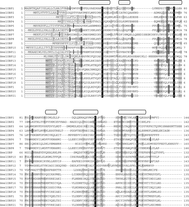
Figure 1.
The alignment of the predicted polypeptides encoding OBPs in Apis mellifera. Conserved residues are highlighted and the signal peptides are in boxes. The rectangular shapes above the alignment represent the α-helices in AmelOBP1 secondary structure. The splice sites are labeled with separators: Vertical ones indicate splice sites between codons; backward slanted separators point out splice sites within codons after the first base.