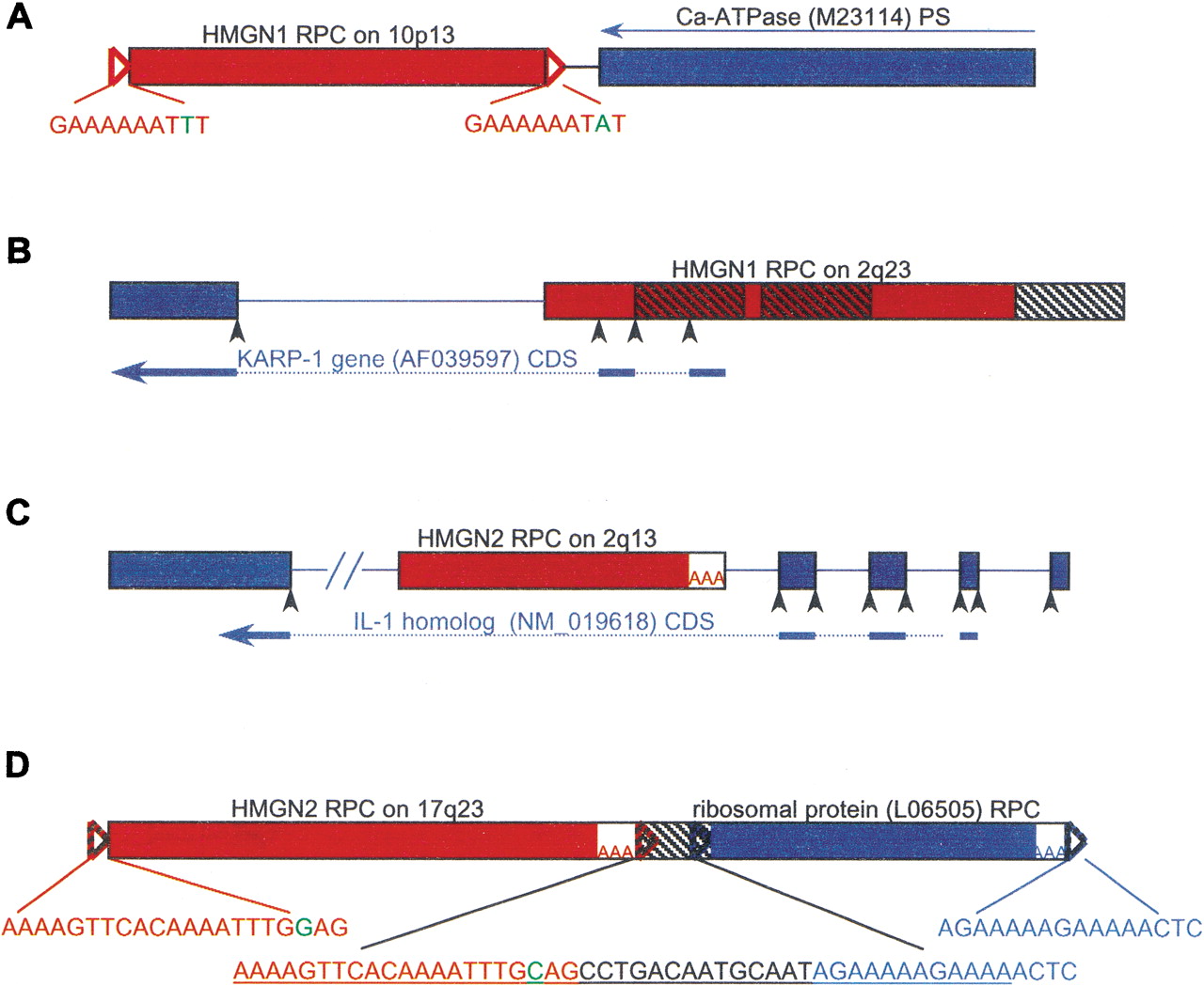
Genes and pseudogenes flanking HMG RPCs. HMG RPC (shown as red boxes) flanking sequences were masked and used in a BLAST search to find surrounding genes and pseudogenes (depicted as blue boxes). The orientation of all HMG RPCs is left to right. (A) A pseudogene of the Ca-ATPase (M23114, 92% identical) was found in the reverse orientation (denoted by a thin blue arrow) to an HMGN1 RPC (89% identical), at a distance of 40 bp (intervening black line). (B) An HMGN1 RPC (88% identical) in the 5′ flank of the KARP-1 gene contributing two exons. The three hatched boxes depict Alu elements. (C) An HMGN2 RPC (89% identical) in the fourth intron of the IL-1 homolog. (D) An HMGN2 RPC (93% identical) upstream and in the same orientation as an RPC of ribosomal protein L12 (95% identical; see text). The 47 bp between them is a fragment of a THE1 element (underlined in the middle insert), from which the TSDs for both RPCs are derived (colored nucleotides). (Open triangles) TSDs; TSD sequences appear in the inserts, and nucleotides in green represent ambiguity; (AAA) poly(A) tracts; (black arrowheads) splice sites; (hatched boxes) repetitive elements; (thick blue arrows) CDS; (dotted lines) position of introns in CDS; (lines between the boxes) intervening genomic DNA, or introns if colored; (–//–) a break artificially inserted into the long sequence for convenient display. Figure not drawn to scale.